. 2018 Aug 8:4:73.
doi: 10.1038/s41394-018-0111-6. eCollection 2018.

Infected charcot spine arthropathy

Affiliations

Infected charcot spine arthropathy

Phani Krishna Karthik Yelamarthy et al. Spinal Cord Ser Cases. .

Abstract

Background: Charcot spinal arthropathy (CSA), a destructive spinal pathology, is seen in patients with impaired sensation. Superimposed infection in the affected spinal segments can lead to a challenge in the diagnosis and management. Spinal cord injury (SCI) is the leading cause of CSA as persons with SCI have significantly impaired sensation. Though infection of the CSA is rare, SCI persons are prone to superimposed infection of the Charcot spine. We report atypical presentations of three cases of CSA with superimposed infection.

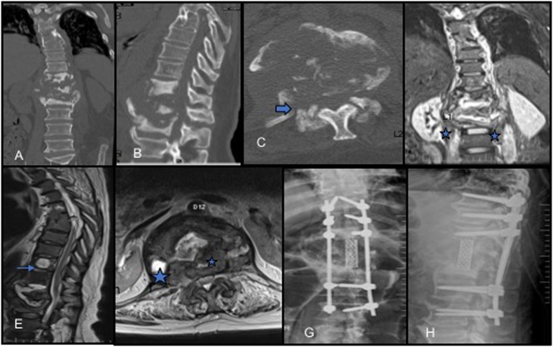
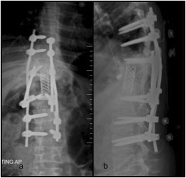
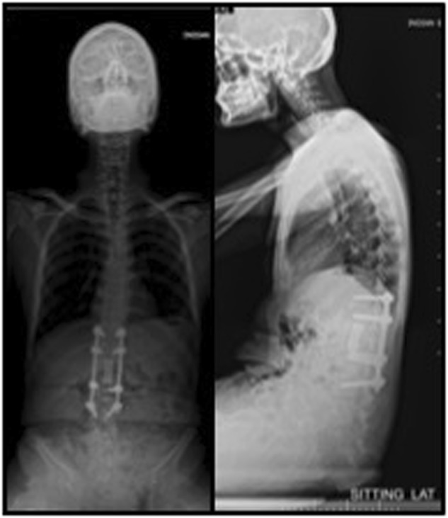
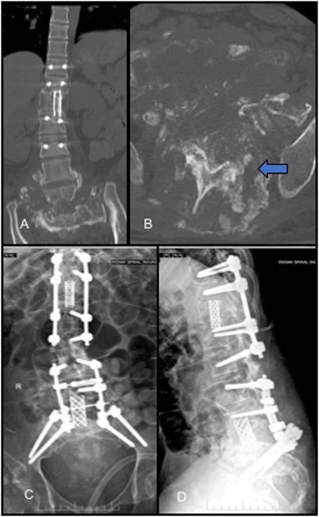
Case descriptions: A 47-year-old male with complete T7-8 SCI developed symptoms suggestive of infection and CSA. He was managed with a posterior vertebral column resection (PVCR) of T12 and intravenous antibiotics as the intraoperative culture showed the growth of E. coli and Pseudomonas. A 26-year-old male with T12 complete paraplegia, post status post open reduction and internal fixation with subsequent implants removal developed infection and CSA over the pseudo-arthrotic lesion with destruction of T12 and L1 vertebrae and an external fistulous track. He was managed with debridement and anterior column T11-L1 reconstruction with a Titanium cage and four-rod pedicle screw stabilization construct. A 25-year-old male with complete paraplegia with CSA at L4-S1. He underwent PVCR of L5 and L3-S2 posterior stabilization. The intraoperative culture and histopathology were suggestive of tuberculous infection.

Conclusion: Pyogenic or tubercular infection of CSA should be considered as a diagnostic possibility in persons with SCI who are more prone to infections. The management includes aggressive debridement and circumferential fusion along with appropriate medications to control the infection.


Conflict of interest statement

Compliance with ethical standardsThe authors declare that they have no conflict of interest.

Figures

Fig. 1
Fig. 1
Preoperative CT (a, b, c) and MRI (d, e) showing gross destruction of L1 vertebral body with associated sclerosis in T12 and L2, facet fragmentation (block arrow; image c), psoas abscess (★; image d, f), and intervertebral fluid (small blue arrow; image e). Six month postoperative antero-posterior and lateral radiographs showing a stable construct (Image g, h)
Fig. 2
Fig. 2
Preoperative radiographs (a, b), CT (c, d, e), and MRI (f, g, h) showing partial destruction of T11 and T12 vertebrae with associated sclerosis, facet fragmentation (block arrow; image e), fistulous track (curved arrow; image g), and syrinx (small arrow, image h)
Fig. 3
Fig. 3
One year postoperative antero-posterior and lateral radiographs with four rods and anterior mesh cage
Fig. 4
Fig. 4
Preoperative radiographs (a, b), showing previous T11–L3 fusion with destruction of the lumbo-sacral junction
Fig. 5
Fig. 5
CT scan showing (coronal and axial images-Image a, b) showing near total destruction of L5 vertebra with facet fragmentation (block arrow; image b) but no accompanying sclerosis. Postoperative antero-posterior and lateral radiographs (Image c, d)

References

    1. Barrey C, Massourides H, Cotton F, Perrin G, Rode G. Charcot spine: two new case reports and a systematic review of 109 clinical cases from the literature. Ann Phys Rehabil Med. 2010;53:200–20. doi: 10.1016/j.rehab.2009.11.008. - DOI - PubMed
    1. Moreau S, Lonjon G, Jameson R, Judet T, de Loubresse CG. Do all charcot spine require surgery? Orthop Traumatol Surg Res. 2014;100:779–84. doi: 10.1016/j.otsr.2014.05.021. - DOI - PubMed
    1. Ledbetter LN, Salzman KL, Sanders RK, Shah LM. Spinal neuroarthropathy: pathophysiology, clinical and imaging features, and differential diagnosis. Radiographics. 2016;36:783–99. doi: 10.1148/rg.2016150121. - DOI - PubMed
    1. Jacobs WB, Bransford RJ, Bellabarba C, Chapman JR. Surgical management of Charcot spinal arthropathy: a single-center retrospective series highlighting the evolution of management. J Neurosurg Spine. 2012;17:422–31. doi: 10.3171/2012.7.SPINE111039. - DOI - PubMed
    1. Capps E, Linnau KF, Crane DA. Beyond broken spines–what the radiologist needs to know about late complications of spinal cord injury. Insights Imaging. 2015;6:111–22. doi: 10.1007/s13244-014-0375-8. - DOI - PMC - PubMed

LinkOut - more resources