

Characterisation of the Morphological, Functional and Molecular Changes in Sunitinib-Resistant Renal Cell Carcinoma Cells
- PMID: 30109169
- PMCID: PMC6088203
- DOI: 10.15586/jkcvhl.2018.106
Characterisation of the Morphological, Functional and Molecular Changes in Sunitinib-Resistant Renal Cell Carcinoma Cells
Abstract
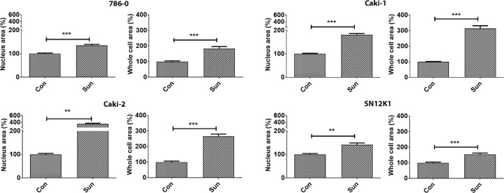
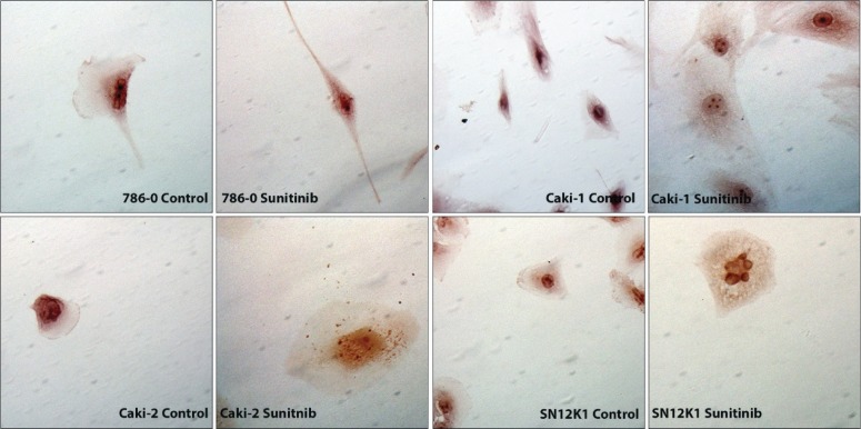
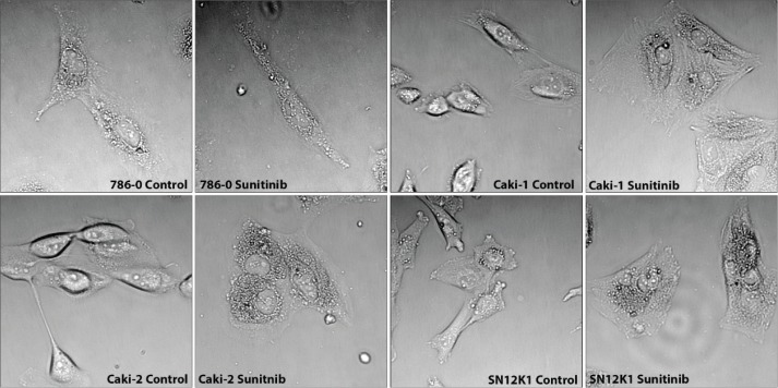
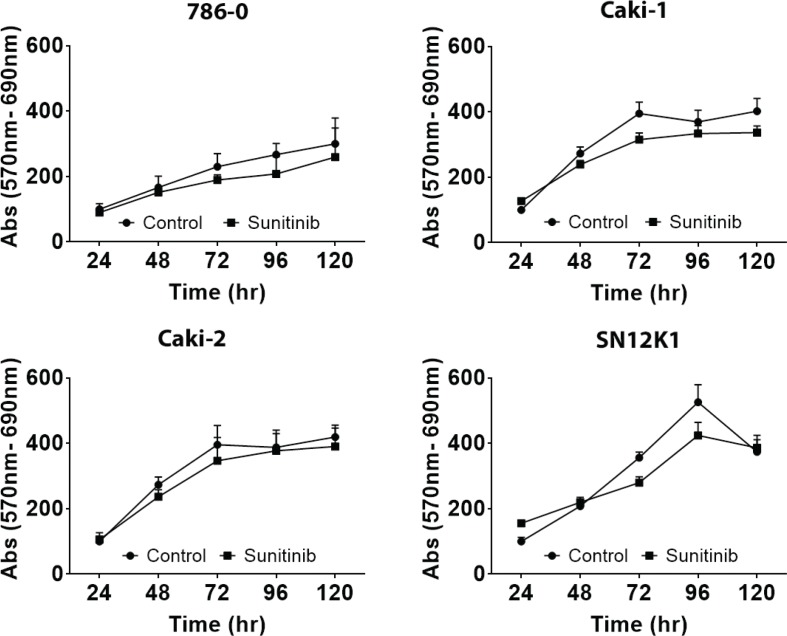
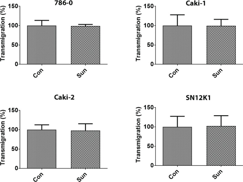
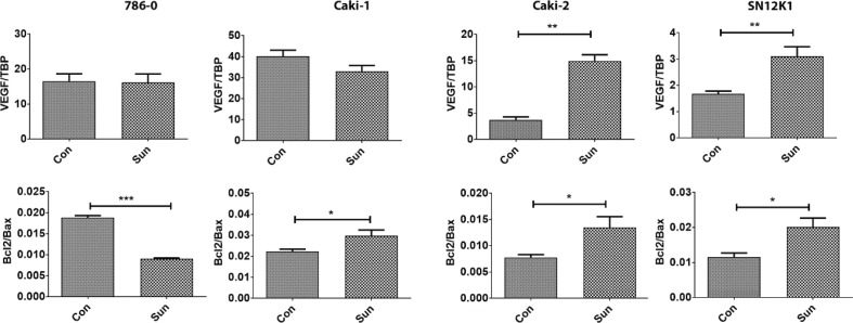
Sunitinib resistance is a major clinical problem hampering the treatment of renal cell carcinoma (RCC). Studies on the comprehensive characterisation of morphological, functional and molecular changes in sunitinib-resistant RCC cells are lacking. The aim of the current study was to develop sunitinib resistance in four human RCC cell lines (786-0, Caki-1, Caki-2 and SN12K1), and to characterise the changed cell biology with sunitinib resistance. RCC cells were made resistant by continuous, chronic exposure to 10 μM of sunitinib over a period of 12 months. Cell proliferation, morphology, transmigration, and gene expression for interleukin-6 (IL-6), interleukin-8 (IL-8), vascular endothelial growth factor (VEGF), Bcl-2 and Bax were studied. There was no significant difference in growth rate or transmigration between the parental and resistant cells. Sunitinib-resistant cells were significantly hypertrophic compared with parental cells as evidenced by increases in the surface areas of the whole cells and the nuclei. IL-6 was significantly increased in all resistant cells. IL-8 was increased in sunitinib-resistant Caki-2 and SN12K1 cells and decreased in 786-0 without any significant changes in Caki-1. VEGF was increased in resistant Caki-2 and SN12K1 cells but not in 786-0 and Caki-1. The Bcl2/Bax ratio was increased in Caki-1, Caki-2 and SN12K1 cells but decreased in 786-0 cells. The increased IL-6 may contribute to sunitinib resistance either via VEGF-mediated angiogenesis or through shifting of the Bcl2/Bax balance in favour of anti-apoptosis.
Keywords: angiogenesis; anti-apoptosis; interleukin-6; renal cell carcinoma; sunitinib resistance.
Figures

Similar articles
-
Cellular Adaptation to VEGF-Targeted Antiangiogenic Therapy Induces Evasive Resistance by Overproduction of Alternative Endothelial Cell Growth Factors in Renal Cell Carcinoma.Neoplasia. 2015 Nov;17(11):805-16. doi: 10.1016/j.neo.2015.11.001. Neoplasia. 2015. PMID: 26678908 Free PMC article.
-
Decreased apoptosis repressor with caspase recruitment domain confers resistance to sunitinib in renal cell carcinoma through alternate angiogenesis pathways.Biochem Biophys Res Commun. 2016 Apr 22;473(1):47-53. doi: 10.1016/j.bbrc.2016.03.048. Epub 2016 Mar 17. Biochem Biophys Res Commun. 2016. PMID: 26995091
-
NGAL can alternately mediate sunitinib resistance in renal cell carcinoma.J Urol. 2014 Aug;192(2):559-66. doi: 10.1016/j.juro.2013.12.049. Epub 2014 Jan 11. J Urol. 2014. PMID: 24423438
-
Interleukin-6 induces drug resistance in renal cell carcinoma.Fukushima J Med Sci. 2018 Dec 8;64(3):103-110. doi: 10.5387/fms.2018-15. Epub 2018 Oct 23. Fukushima J Med Sci. 2018. PMID: 30369518 Free PMC article. Review.
-
Resistance to sunitinib in renal cell carcinoma: From molecular mechanisms to predictive markers and future perspectives.Biochim Biophys Acta. 2015 Jan;1855(1):1-16. doi: 10.1016/j.bbcan.2014.11.002. Epub 2014 Nov 11. Biochim Biophys Acta. 2015. PMID: 25446042 Review.
Cited by
-
Protein kinase CK2 sustains de novo fatty acid synthesis by regulating the expression of SCD-1 in human renal cancer cells.Cancer Cell Int. 2024 Dec 26;24(1):432. doi: 10.1186/s12935-024-03611-y. Cancer Cell Int. 2024. PMID: 39726006 Free PMC article.
-
Identification of low-dose multidrug combinations for sunitinib-naive and pre-treated renal cell carcinoma.Br J Cancer. 2020 Aug;123(4):556-567. doi: 10.1038/s41416-020-0890-y. Epub 2020 May 22. Br J Cancer. 2020. PMID: 32439932 Free PMC article.
-
Prognostic significance of VHL, HIF1A, HIF2A, VEGFA and p53 expression in patients with clear‑cell renal cell carcinoma treated with sunitinib as first‑line treatment.Int J Oncol. 2019 Aug;55(2):371-390. doi: 10.3892/ijo.2019.4830. Epub 2019 Jun 25. Int J Oncol. 2019. PMID: 31268155 Free PMC article.
-
Efficacy and Prognostic Factors of Sunitinib as First-Line Therapy for Patients With Metastatic Renal Cell Carcinoma in an Arab Population.JCO Glob Oncol. 2020 Feb;6:19-26. doi: 10.1200/JGO.19.00111. JCO Glob Oncol. 2020. PMID: 32031432 Free PMC article. Review.
-
Integrated mRNA and miRNA Transcriptomic Analyses Reveals Divergent Mechanisms of Sunitinib Resistance in Clear Cell Renal Cell Carcinoma (ccRCC).Cancers (Basel). 2021 Aug 31;13(17):4401. doi: 10.3390/cancers13174401. Cancers (Basel). 2021. PMID: 34503211 Free PMC article.
References
LinkOut - more resources
Full Text Sources
Other Literature Sources
Research Materials