Review
. 2018 Nov;27(11):1876-1892.
doi: 10.1002/pro.3496.

The nuclear receptor superfamily: A structural perspective

Affiliations
Review

The nuclear receptor superfamily: A structural perspective

Emily R Weikum et al. Protein Sci. 2018 Nov.

Abstract

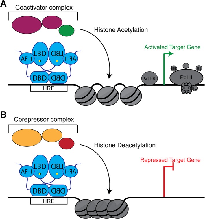
Nuclear receptors (NRs) are a family of transcription factors that regulate numerous physiological processes such as metabolism, reproduction, inflammation, as well as the circadian rhythm. NRs sense changes in lipid metabolite levels to drive differential gene expression, producing distinct physiologic effects. This is an allosteric process whereby binding a cognate ligand and specific DNA sequences drives the recruitment of diverse transcriptional co-regulators at chromatin and ultimately transactivation or transrepression of target genes. Dysregulation of NR signaling leads to various malignances, metabolic disorders, and inflammatory disease. Given their important role in physiology and ability to respond to small lipophilic ligands, NRs have emerged as valuable therapeutic targets. Here, we summarize and discuss the recent progress on understanding the complex mechanism of action of NRs, primarily from a structural perspective. Finally, we suggest future studies to improve our understanding of NR signaling and better design drugs by integrating multiple structural and biophysical approaches.

Keywords: DNA binding domain; co-regulator; ligand binding domain; nuclear receptor; transactivation; transrepression.


Figures

Figure 1
Figure 1
Modular domain structure of NRs. (A) Basic modular domain structure of NRs is composed of an unstructured NTD that contains the Activation Function 1 (AF‐1) surface, a zinc finger DBD, a flexible hinge region, and a LBD that binds to ligands and interacts with co‐regulator proteins through the Activation Function 2 (AF‐2) surface. (B) General domain size and amino acid length of a variety of NRs. The DBD and LBDs are the most conserved regions where as the other domains are more variable in length and sequence composition. (C) Example of a full‐length NR structure shows LXR‐RXR heterodimer (PDB: http://firstglance.jmol.org/fg.htm?mol=4NQA) (DBD colored purple, hinge region in yellow, and LBD in green).
Figure 2
Figure 2
NR DNA binding domains. (A) Cartoon representation of NR DBDs indicating important motifs. This domain contains two subdomains, each containing one zinc finger. The first subdomain residues interact with the DNA major groove to make base‐specific interactions on genomic response elements. The second subdomain participates in DBD dimerization and makes non‐specific contacts with the DNA backbone. Some NRs, like LRH‐1 and GCNF, also contain C‐terminal extensions (CTEs) that make base‐specific contacts with the minor groove. (B) Cartoon representation of folded GR DBD highlighting the important regions (PDB: http://firstglance.jmol.org/fg.htm?mol=3FYL). Zinc atoms are represented as spheres.
Figure 3
Figure 3
NR ligand binding domains. Cartoon representation of the structurally conserved NR LBD. This domain is composed of 11 α‐helices and 4 β‐strands that fold into three layers of a helical sandwich bundle. This fold creates a hydrophobic ligand binding pocket at the bottom of the receptor. This domain also contains the AF surface, composed of H3, H4, and the AF‐H, which interacts with co‐regulator proteins (PDB: http://firstglance.jmol.org/fg.htm?mol=1PZL).
Figure 4
Figure 4
NR ligand interactions. Close up view of SR LBPs showing that (A) GR LBD‐cortisol (PDB: http://firstglance.jmol.org/fg.htm?mol=4P6X) and (B) ER LBD‐estradiol (PDB: http://firstglance.jmol.org/fg.htm?mol=1ERE) use conserved Glu and Arg residues (blue sticks) to make hydrogen bonding interactions (red) with steroid ligands. These interactions help orient the ligand within the pocket. (C) Close up views of FXR LBD‐CDCA (PDB: http://firstglance.jmol.org/fg.htm?mol=1OT7) and (D) LXR LBD‐epoxycholesterol (PDB: http://firstglance.jmol.org/fg.htm?mol=1P8D) show, despite similar ligands, the receptors orient them in opposite directions. This allows natural ligands to discriminate between NRs whose LBDs are highly conserved (E) Comparisons of ligand cavity sizes between GR (PDB: http://firstglance.jmol.org/fg.htm?mol=4P6X), FXR (PDB: http://firstglance.jmol.org/fg.htm?mol=1OT7), and PPAR (PDB: http://firstglance.jmol.org/fg.htm?mol=5AZV).
Figure 5
Figure 5
Genomic response elements. Nuclear receptors bind to genomic response elements (RE) that come in a variety of forms. (A) Members of the SR subfamily bind to palindromic repeats (shown as red DNA cartoon). These repeats are separated by different spacer lengths (shown as yellow DNA cartoon). As examples, the ER DBD – estrogen response elements (ERE) and GR DBD – glucocorticoid response element (GRE) crystal structures are shown. (B) Most other NRs bind to direct repeats, which can also be separated by spacers from 0 to 5 bp. The structures of the RXR‐RAR DBD heterodimer is shown in complex with a DR with 1 bp spacer (DR1) and the VDR homodimer DBD is shown in complex with a DR with 3 bp spacer (DR3). (C) Although rare, some NRs bind to DNA as a monomer to extended half site sequences. Examples include LRH‐1 DBD and SF‐1 DBD (PDBs, from left to right: top row – http://firstglance.jmol.org/fg.htm?mol=4AA6, http://firstglance.jmol.org/fg.htm?mol=1DSZ, and http://firstglance.jmol.org/fg.htm?mol=5L0M; bottom row – http://firstglance.jmol.org/fg.htm?mol=3FYL, http://firstglance.jmol.org/fg.htm?mol=1KB4, and http://firstglance.jmol.org/fg.htm?mol=2FF0).
Figure 6
Figure 6
NR dimerization interfaces. Many NRs utilize the H10/H11 surface to form homodimers or heterodimers. (A) ER LBD – estrogen homodimeric complex shows dimerization occurs between H7, H9, H10/11 (PDB: http://firstglance.jmol.org/fg.htm?mol=1ERE). (B) The LXR‐RXR LBD heterodimer shows a similar dimerization interface (PDB: http://firstglance.jmol.org/fg.htm?mol=1UHL). (C) Unlike the other two, the GR LBD homodimer structure revealed a novel dimerization interface (PDB: http://firstglance.jmol.org/fg.htm?mol=1M2Z). The dimerization interface is colored blue, ligands are shown as sticks (green) and co‐regulator peptides are colored yellow.
Figure 7
Figure 7
NR co‐regulator interactions. (A) Cartoon representation of the co‐regulator LXXLL peptide (green) interacting with the AF surface (purple). The peptide is held in place by a conserved charge clamp interaction formed by a glutamate on H12 and a lysine on H3. (B) Cartoon representation of co‐repressor peptides (pink) interacting with the AF surface (blue). Co‐repressors contain extended (L/I)XX(I/V)I or LXXX(I/L)XXX(I/L) motifs that do not allow for the charge clamp formation. The basis of the “mouse‐trap” model was made by comparing the apo (C) and ligand bound (D) structures of RXR. Upon ligand binding a large rearrangement of H12 is seen (PDBs: http://firstglance.jmol.org/fg.htm?mol=1LBD, http://firstglance.jmol.org/fg.htm?mol=1MVC). (E,F) The more favored “dynamic stabilization” model of NR activation suggests H12 does not undergo such a large conformational change, but instead H12 flexible and ligand binding simply stabilizes the helix. This model was proposed after other apo NR structures, did not show H12 displaced and, upon ligand binding, there was little change in the location of this helix (PDBs: http://firstglance.jmol.org/fg.htm?mol=4DOR, http://firstglance.jmol.org/fg.htm?mol=4PLE). Co‐regulator peptides are colored blue and ligands are shown as sticks (green).
Figure 8
Figure 8
Schematic of NR signaling mechanisms. (A) Type I receptors reside in the cytoplasm (C) in complex with chaperone proteins. Upon ligand binding (hexagon), the receptor is released from this complex and is trafficked into the nucleus (N) where they typically bind to palindromic hormone response elements (HREs) as a homodimer to regulate transcription. (B) Type II receptors are localized in the nucleus. In their unliganded state, they interact with co‐repressor proteins, but upon ligand binding are exchanged for co‐activators. NRs in this group generally form heterodimeric complexes with RXR. (C) Similar to Type II receptors, Type III receptors reside in the nucleus and exchange bound co‐repressors and co‐activators. These receptors bind to direct repeat HREs as homodimers. (D) Type IV receptors are almost identical to Type III except they bind HREs that are extended half sites as monomers.
Figure 9
Figure 9
NRs both activate and repress transcription. (A) To activate gene expression, NRs (blue) interact with their DNA response elements. DNA‐bound NRs recruit co‐activator proteins (magenta), which in turn recruit histone‐modifying enzymes. These histone‐modifying enzymes are commonly histone acetylases (green), which acetylate histone tails. This modification is a mark of active chromatin. Ultimately, the general transcriptional machinery and RNA Polymerase Pol II (gray) are recruited to drive gene expression. (B) To repress transcription, NRs recruit co‐repressor proteins (orange). These proteins recruit other histone deacetylases (red) that reverse histone acetylation and restrict chromatin accessibility. This condensation prevents the transcriptional machinery from accessing the DNA, thus repressing gene expression.

References

    1. Kininis M, Kraus WL (2008) A global view of transcriptional regulation by nuclear receptors: gene expression, factor localization, and DNA sequence analysis. Nucl Receptor Signal 6:e005. - PMC - PubMed
    1. Gustafsson JA (2016) Historical overview of nuclear receptors. J Steroid Biochem Mol Biol 157:3–6. - PubMed
    1. Mangelsdorf DJ, Thummel C, Beato M, Herrlich P, Schutz G, Umesono K, Blumberg B, Kastner P, Mark M, Chambon P, Evans RM (1995) The nuclear receptor superfamily: the second decade. Cell 83:835–839. - PMC - PubMed
    1. Govindan MV, Devic M, Green S, Gronemeyer H, Chambon P (1985) Cloning of the human glucocorticoid receptor cDNA. Nucleic Acids Res 13:8293–8304. - PMC - PubMed
    1. Xu EH, Lambert MH (2003) Structural insights into regulation of nuclear receptors by ligands. Nucl Recept Signal 1:e004. - PMC - PubMed

Publication types

LinkOut - more resources