. 2019 Apr;16(4):504-517.
doi: 10.1080/15476286.2018.1504546. Epub 2018 Sep 18.

PAM identification by CRISPR-Cas effector complexes: diversified mechanisms and structures

Affiliations

PAM identification by CRISPR-Cas effector complexes: diversified mechanisms and structures

Daniel Gleditzsch et al. RNA Biol. 2019 Apr.

Abstract

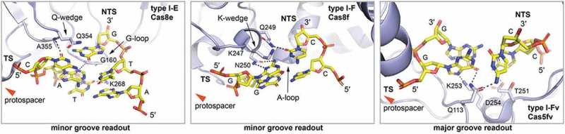
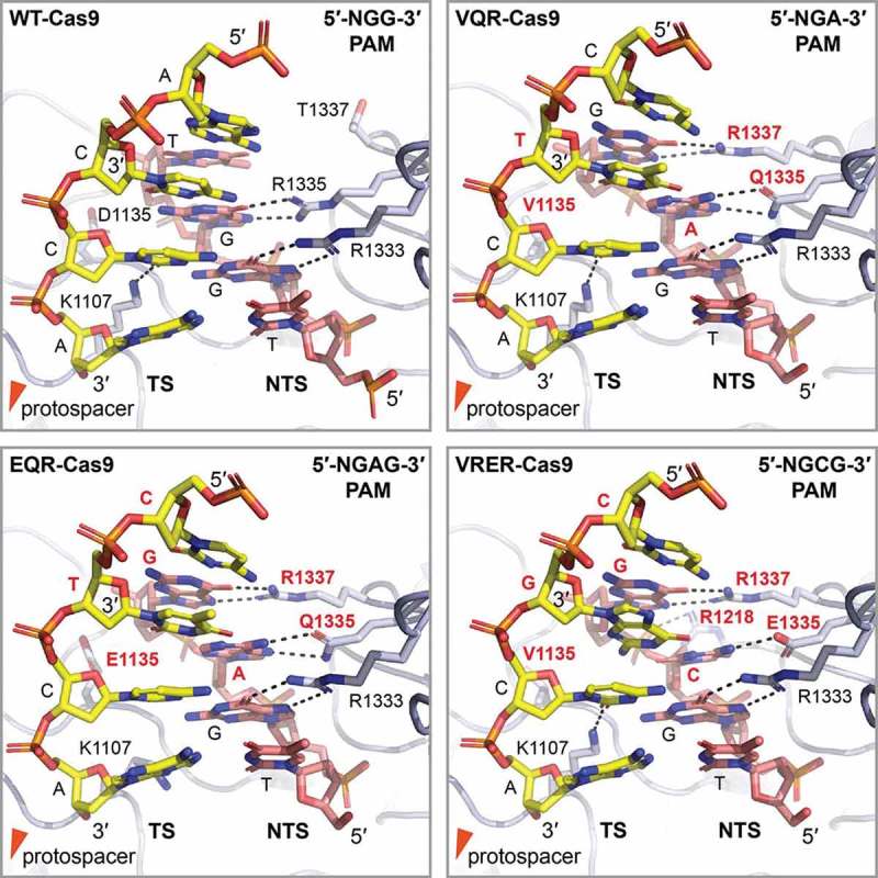
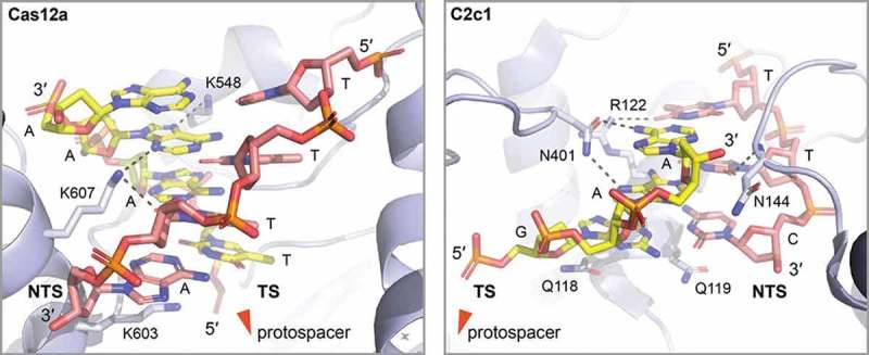
Adaptive immunity of prokaryotes is mediated by CRISPR-Cas systems that employ a large variety of Cas protein effectors to identify and destroy foreign genetic material. The different targeting mechanisms of Cas proteins rely on the proper protection of the host genome sequence while allowing for efficient detection of target sequences, termed protospacers. A short DNA sequence, the protospacer-adjacent motif (PAM), is frequently used to mark proper target sites. Cas proteins have evolved a multitude of PAM-interacting domains, which enables them to cope with viral anti-CRISPR measures that alter the sequence or accessibility of PAM elements. In this review, we summarize known PAM recognition strategies for all CRISPR-Cas types. Available structures of target bound Cas protein effector complexes highlight the diversity of mechanisms and domain architectures that are employed to guarantee target specificity.

Keywords: CRISPR; Cas proteins; DNA recognition; PAM; ribonucleoproteins.


Figures

Figure 1.
Figure 1.
PAM recognition by Cas1-Cas2. a: Crystal structure of the E. coli Cas1-Cas2 bound to a dual-forked PAM containing DNA (PDB: 5DQZ [60]). Two copies of Cas2 (light and dark grey) bridge two juxtaposed dimers of Cas1 (light and dark blue). Association of the B-from DNA duplex (orange surface representation) on top of the complex induces bending of the DNA. Cas1a and Cas1b enclose the PAM complementary 3′-overhang (PAM) of the forked DNA. b: Close up on the CTT 3′-overhang (yellow) specific binding pocket formed by Cas1a (light blue) and Cas1b (dark blue). Left: Base specific hydrogen bonding confers specificity (black dotted lines). Right: Surface charge representation of the binding pocket. Cas1a and Cas1b tightly enclose the hook shaped CTT 3′-overhang to provide specificity. Notably, while purine bases would sterically clash with the binding pocket, only pyrimidine bases can be accommodated.
Figure 2.
Figure 2.
PAM recognition by type I Cascades. Left: Close up on the PAM interacting region of the E. coli type I-E Cascade subunit Cas8e (light blue) (PDB: 5H9E [75]). Cas8e promiscuously recognizes the ATG PAM (yellow) via a set of polar interactions (black dashed lines) from the DNA minor groove. The glycine rich loop (G-loop) recognizes the second base pair of the PAM. The Q-wedge might assist in target strand (TS) protospacer displacement from the non-target strand (NTS) protospacer complementary sequence. The red arrow indicates the direction of the protospacer. Middle: Close up on the PAM interacting region of the P. aeruginosa type I-F Cascade subunit Cas8f (light blue) (PDB: 6B44 [38]). Cas8e specifically recognizes the GG PAM (yellow) via a set of polar interactions (black dashed lines) from the DNA minor groove. The alanine rich loop (A-loop) recognizes the second base pair of the PAM. The K-wedge might assist in target strand (TS) protospacer displacement from the non-target strand (NTS) protospacer complementary sequence. The red arrow indicates the direction of the protospacer. Right: Close up on the PAM interacting region of the S. putrefaciens type I-Fv Cascade subunit Cas5fv (light blue) (PDB: 5O6U [82]). Cas5fv specifically recognizes the GG PAM (yellow) via a set of polar interactions (black dashed lines) from the DNA major groove. Base pairing of the first PAM GC base pair is distorted by Q113. The red arrow indicates the direction of the protospacer.
Figure 3.
Figure 3.
PAM recognition by wildtype and PAM specificity engineered Cas9 variants. Shown is the detailed view on the PAM interacting region of wildtype Cas9 (PDB: 4UN3 [98]) and the three engineered Cas9 versions VQR-Cas9, EQR-Cas9 and VRER-Cas9 (PDB: 5B2R, 5B2S and 5B2T [104]) in the respective panels. Cas9 (light blue) specifically recognizes the GG, NGA, NGAG and NGCG PAM respectively, mainly by polar interactions (black dashed lines) with the non-target strand (NTS, red colored) via the major groove of the PAM containing DNA duplex. K1107, with exception of the EQR-Cas9 variant, forms a hydrogen bond in the minor groove with the target strand (TS) cytosine of the first GC base pair of the PAM, further contributing to specificity. Multiple mutations in the engineered Cas9 variants (red label) result in displacement of the phosphodiester backbone of the PAM duplex and allow the side chain in position 1135 to recognize the altered third PAM nucleotide from the minor groove [104]. Altered PAM nucleotides are labeled red for clarity.
Figure 4.
Figure 4.
PAM recognition by type V Cas12a and C2c1. Left: Close up on the PAM interacting region of the Acidaminococcus sp. type V Cas12a (light blue) (PDB: 5B43 [117]). Cas12a tightly encloses and recognizes the TTTN PAM by a set of polar interactions from the minor and major groove of the PAM containing duplex. For clarity, van der Waals interaction mediating side chains are not shown. Notably, Lysine K603 stacks under the last nucleotide of the non-target strand (NTS, red), potentially assisting in target (TS, yellow) and non-target strand separation. Right: Close up on the PAM interacting region of the Alicyclobacillus acidoterrestris type V C2c1 (light blue) (PDB: 5U30 [116]). C2c1 tightly encloses and recognizes the TTC PAM by a set of polar interactions from the minor and major groove of the PAM containing duplex. For clarity, van der Waals interaction mediating side chains are not shown. Noteworthy, two glutamines (Q118 and Q119) stack under the last PAM base pair potentially assisting in target (TS, yellow) and non-target strand (NTS, red) separation.

References

    1. Barrangou R, Fremaux C, Deveau H, et al. CRISPR provides acquired resistance against viruses in prokaryotes. Science. 2007. March 23;315(5819):1709–1712. PubMed PMID: 17379808. - PubMed
    1. Brouns SJ, Jore MM, Lundgren M, et al. Small CRISPR RNAs guide antiviral defense in prokaryotes. Science. 2008;321(5891):960–964. - PMC - PubMed
    1. Nunez JK, Kranzusch PJ, Noeske J, et al. Cas1-Cas2 complex formation mediates spacer acquisition during CRISPR-Cas adaptive immunity. Nat Struct Mol Biol. 2014. June;21(6):528–534. PubMed PMID: 24793649; PubMed Central PMCID: PMC4075942. - PMC - PubMed
    1. Richter C, Dy RL, McKenzie RE, et al. Priming in the Type I-F CRISPR-Cas system triggers strand-independent spacer acquisition, bi-directionally from the primed protospacer. Nucleic Acids Res. 2014. July;42(13):8516–8526. PubMed PMID: 24990370; PubMed Central PMCID: PMC4117759. - PMC - PubMed
    1. Nunez JK, Lee AS, Engelman A, et al. Integrase-mediated spacer acquisition during CRISPR-Cas adaptive immunity. Nature. 2015. March 12;519(7542):193–198. PubMed PMID: 25707795; PubMed Central PMCID: PMCPMC4359072. - PMC - PubMed

Publication types