

Review
doi: 10.1152/physiol.00013.2018.
The Neurobiological Basis of Sleep and Sleep Disorders
Affiliations
- PMID: 30109824
- PMCID: PMC6230548
- DOI: 10.1152/physiol.00013.2018
Item in Clipboard
Review
The Neurobiological Basis of Sleep and Sleep Disorders
Physiology (Bethesda).
.
Abstract
The functions of sleep remain a mystery. Yet they must be important since sleep is highly conserved, and its chronic disruption is associated with various metabolic, psychiatric, and neurodegenerative disorders. This review will cover our evolving understanding of the mechanisms by which sleep is controlled and the complex relationship between sleep and disease states.
Figures
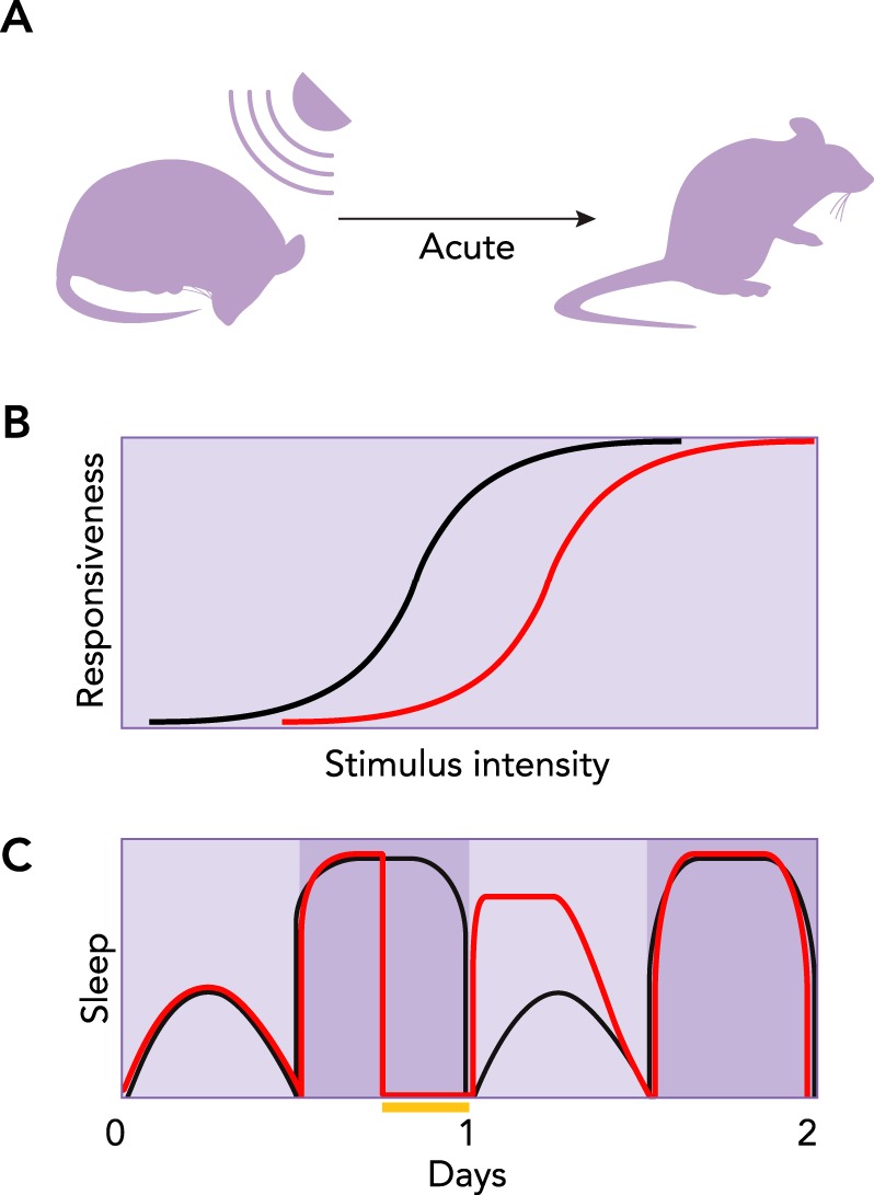
Core behavioral features of sleep A: rapidly reversible quiescence can be demonstrated with a sufficiently strong stimulus, thus distinguishing sleep from seizure, coma, hibernation, and anesthesia. B: reduced arousal. A greater stimulus intensity is required to evoke a given behavioral response during sleep (red line) than during wakefulness (black line). C: the homeostatic nature of sleep is apparent from animals’ attempts to compensate for lost sleep. Black and red lines illustrate typical sleep cycles for control and sleep-deprived groups, respectively. Yellow bar indicates period of sleep deprivation. The next day, the sleep-deprived group sleeps more (i.e., rebounds) compared with the control.
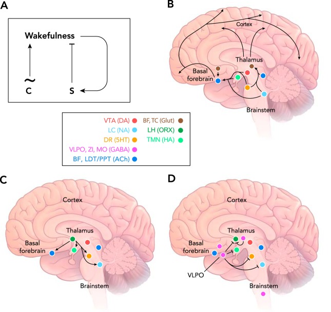
Neuroanatomy of sleep control A: two process model for control of the sleep/wake cycle by the circadian clock (process C) and the sleep homeostat (process S). The clock drives a cycling pattern of arousal with a period of ~24 h. Process S increases during waking until it exceeds a certain threshold, after which it discharges, leading to suppression of waking and initiation of sleep. B: wake-promoting loci in the mammalian brain, including neuromodulatory centers of the ascending reticular activating system. C: orexin neurons in the lateral hypothalamus excite wake-promoting loci to stabilize the waking state. D: GABAergic inhibition of wake-promoting loci and other unknown targets promotes sleep. Color-coded key: BF, basal forebrain; TC, thalamocortical relay neurons; LH, lateral hypothalamus; VTA, ventral tegmental area; LC, locus coeruleus; DR, dorsal raphe nucleus; VLPO, ventrolateral preoptic nucleus; ZI, zona inserta of the subthalamus; MO, medulla oblongata; LDT/PPT, laterodorsaltegmental and pedunculopontine nuclei. Glut, glutamate; ORX, orexin; HA, histamine; DA, dopamine; NA, noradrenaline; 5HT, serotonin; ACh, acetylcholine.
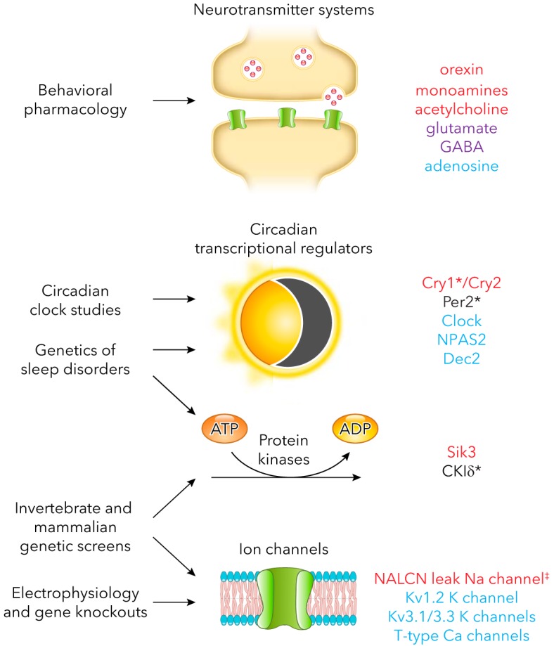
Endogenous molecules that impact sleep and the approaches that led to their discovery Only mammalian molecules are listed. Red, promotes waking; blue, promotes sleep; purple, promotes waking and sleep; asterisk, regulates sleep timing. Not listed: components of metabolic pathways for neurotransmitters, neurotransmitter receptors, and transporters. ‡A gain-of-function mutation in NALCN decreases REM sleep but has no effect on total sleep duration (44). Loss-of-function mutations in the fly ortholog of NALCN, called narrow abdomen, increase sleep (64). Therefore, NALCN has been provisionally assigned a wake-promoting function.
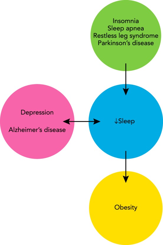
Interaction between sleep and disorders of the nervous system Many disorders disrupt sleep (arrow from green to blue circle). In other cases, low sleep exacerbates existing health problems (arrow from blue to yellow circle). Positive feedback may exist between poor-quality sleep and still other disorders (bidirectional arrow between blue and pink circles).
References
-
- American Psychiatric Association., and American Psychiatric Association. DSM-5 Task Force Diagnostic and Statistical Manual of Mental Disorders: DSM-5. Washington, D.C.: American Psychiatric Association, 2013, p. xliv.
Publication types
MeSH terms
Grants and funding
LinkOut - more resources
Full Text Sources
Other Literature Sources
Medical