Review
. 2018 Aug;8(1):10.1128/ecosalplus.ESP-0004-2018.
doi: 10.1128/ecosalplus.ESP-0004-2018.

An Introduction to the Structure and Function of the Catalytic Core Enzyme of Escherichia coli RNA Polymerase

Affiliations
Review

An Introduction to the Structure and Function of the Catalytic Core Enzyme of Escherichia coli RNA Polymerase

Catherine Sutherland et al. EcoSal Plus. 2018 Aug.

Abstract

RNA polymerase (RNAP) is the essential enzyme responsible for transcribing genetic information stored in DNA to RNA. Understanding the structure and function of RNAP is important for those who study basic principles in gene expression, such as the mechanism of transcription and its regulation, as well as translational sciences such as antibiotic development. With over a half-century of investigations, there is a wealth of information available on the structure and function of Escherichia coli RNAP. This review introduces the structural features of E. coli RNAP, organized by subunit, giving information on the function, location, and conservation of these features to early stage investigators who have just started their research of E. coli RNAP.


Figures

Figure 1
Figure 1
Overview of RNAP assembly and structure. (A) Assembly scheme of the RNAP core enzyme. (B) Structural overview of the E. coli RNAP core enzyme shown as a molecular surface representation (α1: yellow, α2: green, β: cyan, β′: pink, and ω: gray) (Protein Data Bank [PDB]: 4YG2). The DNA binding main channel is indicated by a black arrow. Individual subunits are also depicted with partially transparent surface to expose the ribbon model inside. Lineage-specific insertions found in the β (βi4 and βi9) and β′ subunits (β′i6) are indicated in blue. The active site is represented by the catalytic Mg2+ ion (magenta sphere) coordinated in the β′ subunit.
Figure 2
Figure 2
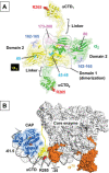
Assembly of alpha dimer and interactions with transcription factors. (A) Dimerization of α subunits (α1: yellow, α2: green). Structural domains and linkers for connecting the αNTD and αCTD are indicated. Amino acid residues contacting with the β and the β′ subunits highlighted in cyan and pink, respectively. Amino acid residues of αNTD responsible for the interaction with CAP are highlighted in blue. Arg265 in the αCTD for the UP element DNA interaction is shown in red color CPK representation. (B) Structural model of the CAP (light blue), RNAP (core enzyme, white; σ70, orange) and lac promoter DNA complex (PDB: 6B6H). The CAP binds DNA centered at position –61.5 relative to the transcription start site and interacts with one copy of the αCTD positioned between the CAP and σ70. A linker (residues 236 to 247) connecting between the NTD and CTD of α subunit is shown as a yellow line.
Figure 3
Figure 3
Interactions of the omega subunit with the β′ subunit and ppGpp. (A) Interaction between the ω subunit and the DPBB domain of β′ subunit. The Mg2+ coordinated at the active site of RNAP is shown as a magenta sphere. (B) ppGpp site 1 (ω, gray; β′, pink and ppGpp, CPK model). Amino acid residues that interact with ppGpp are shown as stick models and indicated. (C) Chemical structure of ppGpp.
Figure 4
Figure 4
Structural features of E. coli RNAP involved in transcription. (A) Structural model of the E. coli RNAP elongation complex (PDB: 6ALF with modification). The core enzyme is depicted as a partially transparent surface model with the DPBB domains of β and β′ subunits (ribbon models). DNA and RNA are shown as CPK models (RNA, red; template DNA, black; nontemplate DNA, light gray). The DNA binding main channel is indicated by a black arrow. (B) Important structural features of the active site (BH, bridge helix; TL, trigger loop). (C) (Top) Rifampin binding pocket of the β subunit (Rifampin, CPK model; β subunit, partially transparent surface with ribbon model). Amino acid residues in the RRDR are highlighted in red. (Bottom) Sequence alignment spanning RRDRs of the E. coli, Thermus thermophilus, and M. tuberculosis RNAP. Amino acids that are identical among the three species are shown as gray background. Three most clinically important RMP resistance mutations are indicated.

References

    1. Feklístov A, Sharon BD, Darst SA, Gross CA. 2014. Bacterial sigma factors: a historical, structural, and genomic perspective. Annu Rev Microbiol 68:357–376. [PubMed] - PubMed
    1. Riordan JT, Mitra A. 2017. Regulation of Escherichia coli pathogenesis by alternative sigma factor N. Ecosal Plus 7:7. [PubMed] - PMC - PubMed
    1. Hurwitz J. 2005. The discovery of RNA polymerase. J Biol Chem 280:42477–42485. [PubMed] - PubMed
    1. Fujita N, Ishihama A. 1996. Reconstitution of RNA polymerase. Methods Enzymol 273:121–130. - PubMed
    1. Artsimovitch I, Svetlov V, Murakami KS, Landick R. 2003. Co-overexpression of Escherichia coli RNA polymerase subunits allows isolation and analysis of mutant enzymes lacking lineage-specific sequence insertions. J Biol Chem 278:12344–12355. [PubMed] - PubMed

Publication types