Guideline
. 2018 Aug 15;198(4):e15-e43.
doi: 10.1164/rccm.201806-1160ST.

Assessment of Right Ventricular Function in the Research Setting: Knowledge Gaps and Pathways Forward. An Official American Thoracic Society Research Statement

Guideline

Assessment of Right Ventricular Function in the Research Setting: Knowledge Gaps and Pathways Forward. An Official American Thoracic Society Research Statement

Tim Lahm et al. Am J Respir Crit Care Med. .

Abstract

Background: Right ventricular (RV) adaptation to acute and chronic pulmonary hypertensive syndromes is a significant determinant of short- and long-term outcomes. Although remarkable progress has been made in the understanding of RV function and failure since the meeting of the NIH Working Group on Cellular and Molecular Mechanisms of Right Heart Failure in 2005, significant gaps remain at many levels in the understanding of cellular and molecular mechanisms of RV responses to pressure and volume overload, in the validation of diagnostic modalities, and in the development of evidence-based therapies.

Methods: A multidisciplinary working group of 20 international experts from the American Thoracic Society Assemblies on Pulmonary Circulation and Critical Care, as well as external content experts, reviewed the literature, identified important knowledge gaps, and provided recommendations.

Results: This document reviews the knowledge in the field of RV failure, identifies and prioritizes the most pertinent research gaps, and provides a prioritized pathway for addressing these preclinical and clinical questions. The group identified knowledge gaps and research opportunities in three major topic areas: 1) optimizing the methodology to assess RV function in acute and chronic conditions in preclinical models, human studies, and clinical trials; 2) analyzing advanced RV hemodynamic parameters at rest and in response to exercise; and 3) deciphering the underlying molecular and pathogenic mechanisms of RV function and failure in diverse pulmonary hypertension syndromes.

Conclusions: This statement provides a roadmap to further advance the state of knowledge, with the ultimate goal of developing RV-targeted therapies for patients with RV failure of any etiology.

Keywords: acute respiratory distress syndrome; pulmonary circulation; pulmonary embolism; pulmonary hypertension; right ventricle.


Figures
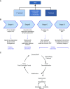
Figure 1.
Figure 1.
Etiologies and stages of right ventricular failure (RVF). (A) RVF occurs as a consequence of alterations in preload, changes in mechanics and/or decreases in contractility, or increases in afterload. (B) Classification of RVF according to stages of development in keeping with the recent classification of the American College of Cardiology Foundation/American Heart Association Task Force for Heart Failure (298, 299). Note that stages are not static and that stages C and D are potentially reversible with normalization or significant decrease in pulmonary vascular resistance (e.g., after pulmonary endarterectomy or lung transplantation) (300, 301). Symptomatic RVF (stage C) is usually managed pharmacologically, whereas refractory RVF (stage D) often requires specialized interventional or surgical measures. Preventive measures may be applied at any of the different stages of RVF. Decompensation may occur at any stage. (C) Cycle of acute on chronic decompensation of RVF. Acute decompensation is usually provoked by one or more precipitating factors (e.g., infection, pulmonary embolism, bleeding disorders) or by progression of the underlying disease (112). Patients frequently deteriorate and enter a vicious cycle of hypotension, ischemia, and decreased pump function or may stabilize and revert to more or less stable chronic RVF. However, mortality after hospital discharge remains high (35% at 12 mo [112]). PAH = pulmonary arterial hypertension; RHF = right heart failure.
Figure 2.
Figure 2.
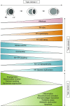
Classifiers and time course of right ventricular failure (RVF). (A) RV function is described using several characteristics, such as stage and acuity, as well as functional class (either by New York Heart Association [NYHA] classification or by cardiopulmonary exercise testing) and severity (using hemodynamic characteristics, markers of RV function, ventriculoatrial [VA] coupling, imaging or plasma biomarkers indicating RV remodeling, as well as presence or absence of arrhythmias [e.g., atrial fibrillation] and presence or absence of systemic involvement [e.g., liver congestion, renal impairment]) (2, 128, 162). Response to therapy (e.g., treatment-responsive vs. refractory RVF) is frequently used for classifying RV function as well. Novel classifiers and “deep” phenotyping tools include imaging approaches aimed at assessing perfusion, metabolism, and/or fibrosis, complemented by “omics” and “big data” approaches. (B) Time course of RVF development in the setting of chronic pressure overload from pulmonary vascular disease. See Reference for detailed review of the supporting literature. Note that significant molecular and temporal heterogeneity exist in RV adaptation and maladaptation to pressure overload, possibly determined by genetic, genomic, epigenetic, and molecular changes due to endogenous and exogenous factors. For example, in scleroderma-associated pulmonary arterial hypertension (PAH), end-systolic elastance (Ees) is lower than in idiopathic PAH, and Ees/arterial elastance (Ea) decreases earlier than in other disease states (170). Cardiac output/cardiac index decreases as a result of transition from adaptive to maladaptive remodeling, because of either progression of the underlying disease or an additional insult. BNP = brain natriuretic peptide; MPAP = mean pulmonary arterial pressure; PVR = pulmonary vascular resistance. (B) Modified from Reference .
Figure 3.
Figure 3.
Concept figure of changes in right ventricular (RV) structural, functional, and biochemical/molecular processes as the RV transitions from normal function to failure in the setting of increased afterload. Afterload increases can be acute (e.g., in cases of pulmonary embolism or acute respiratory distress syndrome) or chronic (e.g., in pulmonary arterial hypertension, pulmonary hypertension from chronic heart or lung disease, or chronic thromboembolic pulmonary hypertension). With progressive increases in RV afterload (top), RV size increases. In forms of chronic afterload increases, RV hypertrophy develops. RV stroke volume, RV contractility, and RV–pulmonary artery (PA) coupling are initially maintained (or even increased) but then progressively decrease. In parallel, RV diastolic dysfunction and RV–left ventricular (LV) dyssynchrony develop. At a molecular/biochemical level, these processes are accompanied by alterations in angiogenesis, calcium handling, mitochondrial function, and sarcomere organization, as well as progressive increases in inflammation, neurohormonal activation, fibrosis/collagen deposition, oxidative stress, metabolic dysfunction, and cardiomyocyte apoptosis. Note that for didactic purposes, long-term trajectories for all changes are shown as gradual increases or decreases, whereas in reality these changes may exhibit fluctuations and not develop in a parallel manner. Topic domain 1 of this research statement focuses on optimizing the methodology to assess RV function in acute and chronic conditions in preclinical models, human studies, and clinical trials. Structural and functional RV changes (shown in red, orange, and blue) are covered in topic domain 2; biochemical/molecular processes (in green) are discussed in topic domain 3.
Figure 4.
Figure 4.
Methods used to estimate right ventricle–pulmonary artery (RV–PA) coupling and diastolic stiffness. In both (A) the volume method, and (B) the pressure method, arterial elastance (Ea) is calculated from the ratio of end-systolic pressure (ESP) to stroke volume (SV). End-systolic elastance (Ees) as an approximation of maximum elastance in the volume method is estimated by the ratio of ESP to end-systolic volume (ESV), which results in a simplified Ees/Ea of SV/ESV. In the pressure method, Pmax is estimated from the nonlinear extrapolation of the early systolic and diastolic portions of the RV pressure curve. Ees is the ratio of (Pmax − mPAP) divided by SV, which results in a simplified Ees/Ea of (Pmax/mPAP − 1). (C) The single-beat method calculates Ees as a straight line drawn from Pmax tangent to RV pressure–relative change in volume relationship. The approach relies on an estimate of Pmax determined from the extrapolation of early and late isovolumic portions of an RV pressure curve and synchronized absolute or relative volume measurements. Ees is then defined by a tangent from Pmax to the pressure–volume relationship, and Ea is defined by a line drawn from the Ees point to end-diastolic volume (EDV) (at zero pressure). (D) Diastolic stiffness (β) is calculated by fitting the nonlinear exponential P = α(eVβ − 1) to pressure and volume measured at the beginning of diastole (BDP, ESV) and the end of diastole (EDP, EDV). Adapted by permission from Reference . BDP = beginning diastolic pressure; EDP = end-diastolic pressure; Evol = Ees estimated by the volume method; mPAP = mean pulmonary arterial pressure; Pmax = RV maximum pressure; sRVP = peak systolic RV pressure.

References

    1. Voelkel NF, Quaife RA, Leinwand LA, Barst RJ, McGoon MD, Meldrum DR, et al. National Heart, Lung, and Blood Institute Working Group on Cellular and Molecular Mechanisms of Right Heart Failure. Right ventricular function and failure: report of a National Heart, Lung, and Blood Institute working group on cellular and molecular mechanisms of right heart failure. Circulation. 2006;114:1883–1891. - PubMed
    1. Vonk-Noordegraaf A, Haddad F, Chin KM, Forfia PR, Kawut SM, Lumens J, et al. Right heart adaptation to pulmonary arterial hypertension: physiology and pathobiology. J Am Coll Cardiol. 2013;62:D22–D33. - PubMed
    1. van de Veerdonk MC, Kind T, Marcus JT, Mauritz GJ, Heymans MW, Bogaard HJ, et al. Progressive right ventricular dysfunction in patients with pulmonary arterial hypertension responding to therapy. J Am Coll Cardiol. 2011;58:2511–2519. - PubMed
    1. Meyer P, Filippatos GS, Ahmed MI, Iskandrian AE, Bittner V, Perry GJ, et al. Effects of right ventricular ejection fraction on outcomes in chronic systolic heart failure. Circulation. 2010;121:252–258. - PMC - PubMed
    1. Riedel M, Stanek V, Widimsky J, Prerovsky I. Longterm follow-up of patients with pulmonary thromboembolism: late prognosis and evolution of hemodynamic and respiratory data. Chest. 1982;81:151–158. - PubMed

Publication types