Review
. 2019 Jan/Feb;32(1):25-37.
doi: 10.1089/vim.2018.0073. Epub 2018 Aug 15.

Chemokine CXCL10 and Coronavirus-Induced Neurologic Disease

Affiliations
Review

Chemokine CXCL10 and Coronavirus-Induced Neurologic Disease

Dominic Skinner et al. Viral Immunol. 2019 Jan/Feb.

Abstract

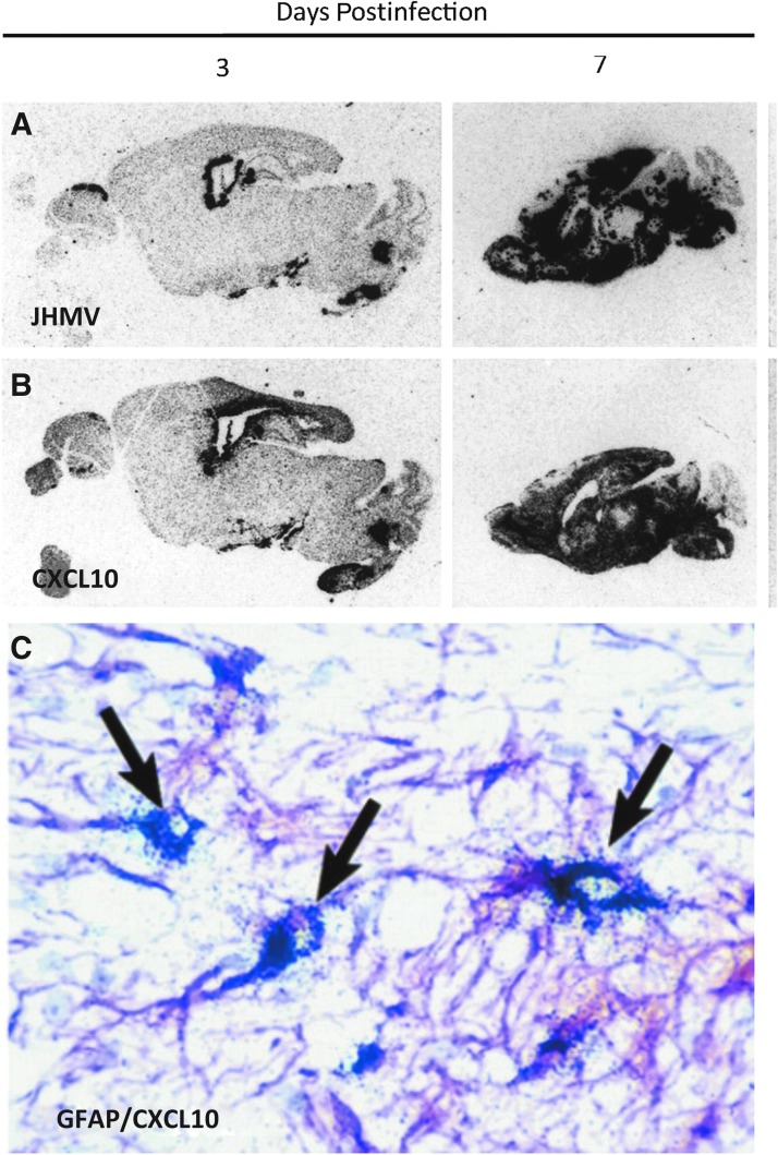
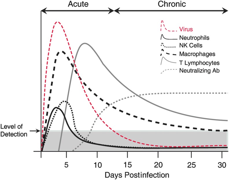
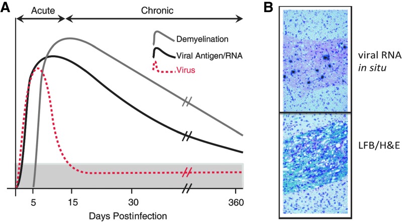
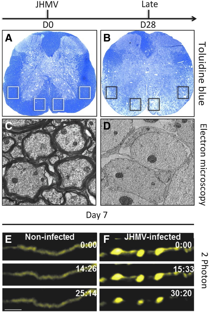
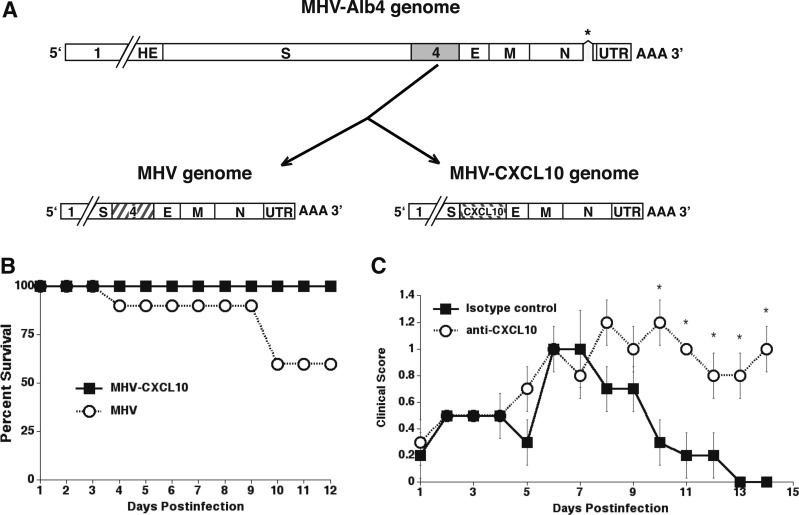
Chemokines (chemotactic cytokines) are involved in a wide variety of biological processes. Following microbial infection, there is often robust chemokine signaling elicited from infected cells, which contributes to both innate and adaptive immune responses that control growth of the invading pathogen. Infection of the central nervous system (CNS) by the neuroadapted John Howard Mueller (JHM) strain of mouse hepatitis virus (JHMV) provides an excellent example of how chemokines aid in host defense as well as contribute to disease. Intracranial inoculation of the CNS of susceptible mice with JHMV results in an acute encephalomyelitis characterized by widespread dissemination of virus throughout the parenchyma. Virus-specific T cells are recruited to the CNS, and control viral replication through release of antiviral cytokines and cytolytic activity. Sterile immunity is not acquired, and virus will persist primarily in white matter tracts leading to chronic neuroinflammation and demyelination. Chemokines are expressed and contribute to defense as well as chronic disease by attracting targeted populations of leukocytes to the CNS. The T cell chemoattractant chemokine CXCL10 (interferon-inducible protein 10 kDa, IP-10) is prominently expressed in both stages of disease, and serves to attract activated T and B lymphocytes expressing CXC chemokine receptor 3 (CXCR3), the receptor for CXCL10. Functional studies that have blocked expression of either CXCL10 or CXCR3 illuminate the important role of this signaling pathway in host defense and neurodegeneration in a model of viral-induced neurologic disease.

Keywords: CNS infection; CXCL10; coronavirus.


Conflict of interest statement

No competing financial interests exist.

Figures

<b>FIG. 1.</b>
FIG. 1.
JHMV infection of the CNS induces rapid expression of CXCL10. In situ hybridization showing distribution of (A) viral RNA and (B) CXCL10 mRNA in brains of MHV-infected mice. Two sequential sagittal sections of paraffin-embedded brain from infected mice at indicated time points were probed with 35S-labeled antisense riboprobes specific to either JHMV or CXCL10. Signal was detected by autoradiography after a 5-day exposure to film. The probes used for each section are indicated. Note the strict colocalization of CXCL10 mRNA with viral RNA at days 2 and 7 postinfection. (C) GFAP-positive astrocyte (purple) cells express CXCL10 mRNA in the CNS of JHMV-infected mice. Combined immunohistochemistry for GFAP and in situ hybridization for CXCL10 mRNA were performed on the brain of a mouse following infection. Astrocytes and their processes are stained purple, and are identified as being positive for CXCL10 mRNA expression at day 7 p.i. by overlaying silver grains (arrows). Original magnification, × 400 (35). CNS, central nervous system; JHMV, John Howard Mueller strain of mouse hepatitis virus; mRNA, messenger RNA; p.i., postinfection.
<b>FIG. 2.</b>
FIG. 2.
JHMV infection of the CNS invokes rapid infiltration of defined immune cell subsets. Cartoon depiction of immune response following i.c. infection of the CNS of susceptible C57BL6 with JHMV. Cellular components of the innate immune response, for example, neutrophils, macrophages, and NK cells are rapidly mobilized, and migrate to the CNS and contribute to opening the blood–brain barrier and controlling viral replication. Infiltrating CD4+ and CD8+ T cells reduce viral titers below level of detection through IFN-γ secretion and cytolytic activity. Neutralizing virus-specific antibody is required to suppress viral recrudescence during chronic disease. i.c., intracranial; NK, natural killer.
<b>FIG. 3.</b>
FIG. 3.
Persistent JHMV infection results in an immune-mediated demyelinating disease. (A) Cartoon depiction of viral persistence within the CNS and demyelination following i.c. infection of C57BL/6 mice with JHMV. Viral titers within the CNS peak between 5 and 7 days p.i., and then decline below levels of detection as a result of infiltrating virus-specific T cells. Sterile immunity is not achieved, and viral RNA/antigen can be detected out to 1 year p.i. Robust immune-mediated demyelination occurs as a result of viral persistence resulting in chronic neuroinflammation. (B) Representative in situ hybridization showing viral RNA (virus-specific 35S-labeled antisense riboprobes) present within a spinal cord white matter tract; sequential spinal cord section stained with LFB/H&E, showing that viral persistence results in immune cell infiltration into white matter tracts accompanied by myelin damage. H&E, hematoxylin and eosin; LFB, luxol fast blue.
<b>FIG. 4.</b>
FIG. 4.
JHMV-induced demyelination and axonal damage. Toluidine blue stained spinal cord sections from (A) control (day 0, D0) and (B) day 28 (D28) postinfection. Demyelination is spread throughout ventral funiculus and lateral white matter columns with notable loss of toluidine blue staining. Electron microscopy reveals extensive loss of myelin sheath at (D) day 28 p.i. compared with (C) noninfected control mice in which thick myelin sheaths are present. Boxed areas in (A, B) indicate regions analyzed for electron microscopic analysis. Focal axonal degeneration occurs in the ventral side of JHMV-infected Thy1-YFP mouse (F) spinal cords when compared with control (E) spinal cords at day 7. 2-Photon time-lapse images (times marked in min:sec) depicting absence of FAD in a noninfected Thy1-YFP spinal cord, scale bars in (E) = 20 μm (21).
<b>FIG. 5.</b>
FIG. 5.
MHV-CXCL10 and MHV have genetic similarity. Both viruses were generated by a recombination reaction with the thermolabile N gene deletion (designated by asterisk) mutant MHV-Alb4 and mRNA generated from a transcription reaction using plasmids that encode from upstream of gene 4 to the 3′ end of MHV-CXCL10 and MHV. (A) The recombination reaction for MHV results in a recombinant that is genetically identical with the WT virus. MHV-CXCL10 is identical with MHV except that gene 4 is replaced by the coding sequence for CXCL10. (B) CXCL10−/− mice i.c. infected with MHV-CXCL10 exhibit 100% survival, whereas only 60% of MHV-infected mice survive to day 12 p.i. (C) Treatment of MHV-CXCL10-infected mice with an anti-CXCL10-neutralizing Ab results in significantly increased (*p ≤ 0.05) clinical scores compared with treatment with an isotype control Ab (data shown are presented as mean ± standard error of the mean) (89). E, E protein (small envelope protein); HE, hemagglutinin-esterase; M, membrane protein; N, nucleocapsid protein; S, surface protein; UTR, 3′ untranslated region; WT, wild type.
<b>FIG. 6.</b>
FIG. 6.
Antibody targeting of CXCL10 in mice persistently infected with JHMV reduces demyelination and increases remyelination. (A) CXCL10 mRNA transcripts were detected by in situ hybridization in white matter tracts of demyelinating spinal cords at day 35 p.i. (A) CXCK10-positive cells (arrows) adjacent to demyelinating lesions. (B) Spinal cord section in which the sense control probe for CRG-2 was used. No positive cells were detected. Original magnification × 400 (35). Toluidine blue-stained transverse section of an (C) anti-CXCL10-treated mouse, showing that the region of demyelination is well defined and limited to the ventral column, whereas in (D) control-treated animals lesions extend throughout the ventral and lateral columns. (E) Electron micrograph of an anti-CXCL10-treated mouse showing axons within the ventral column with thin myelin sheaths (denoted by M and arrow) surrounding axon (a) characteristic of remyelination. (F) Electron micrograph of a control mouse, showing axons (a) within the ventral column with no evidence of remyelination (46).
<b>FIG. 7.</b>
FIG. 7.
CXCL10 treatment results in OPC apoptosis. (A) Secreted CXCL10 protein levels in supernatant from OPC cultures treated with IFN-γ (10, 50, and 100 U/mL—48 h) were measured by ELISA. (B) Treatment of OPC cultures for 6 days with CXCL10 (10 ng/mL) showed a significant increase (*p < 0.05; ***p < 0.0001) in TUNEL positive cells when compared with untreated cultures; values are expressed as mean ± standard deviation. (C) Western blotting of proteins isolated from OPC-enriched cultures obtained from either CXCR3+/+ or CXCR3−/− mice confirms that CXCR3 is expressed in WT cultures. (D) MTT assay showing cell death following 6 days of treatment of CXCR3−/− or WT OPC cultures with either IFN-γ or CXCL10. Cell death is significantly (***p < 0.0001; n = 3 different experiments) reduced in CXCR3−/− cultures compared with WT cultures (82). CXCR3, CXC chemokine receptor 3; ELISA, enzyme-linked immunosorbent assay; OPC, oligodendrocyte progenitor cell.

References

    1. Akwa Y, Hassett DE, Eloranta ML, et al. . Transgenic expression of IFN-alpha in the central nervous system of mice protects against lethal neurotropic viral infection but induces inflammation and neurodegeneration. J Immunol 1998;161:5016–5026 - PubMed
    1. Anghelina D, Zhao J, Trandem K, et al. . Role of regulatory T cells in coronavirus-induced acute encephalitis. Virology 2009;385:358–367 - PMC - PubMed
    1. Baerwald KD, and Popko B. Developing and mature oligodendrocytes respond differently to the immune cytokine interferon-gamma. J Neurosci Res 1998;52:230–239 - PubMed
    1. Balabanov R, Strand K, Kemper A, et al. . Suppressor of cytokine signaling 1 expression protects oligodendrocytes from the deleterious effects of interferon-gamma. J Neurosci 2006;26:5143–5152 - PMC - PubMed
    1. Bender SJ, and Weiss SR. Pathogenesis of murine coronavirus in the central nervous system. J Neuroimmune Pharmacol 2010;5:336–354 - PMC - PubMed

Publication types

MeSH terms