. 2018 Aug 15;14(8):e1007459.
doi: 10.1371/journal.pgen.1007459. eCollection 2018 Aug.

FMRFa receptor stimulated Ca2+ signals alter the activity of flight modulating central dopaminergic neurons in Drosophila melanogaster

Affiliations

FMRFa receptor stimulated Ca2+ signals alter the activity of flight modulating central dopaminergic neurons in Drosophila melanogaster

Preethi Ravi et al. PLoS Genet. .

Abstract

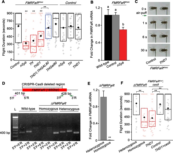
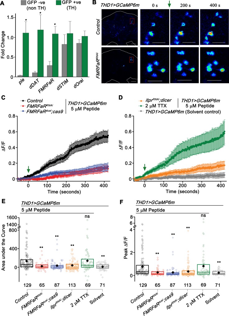
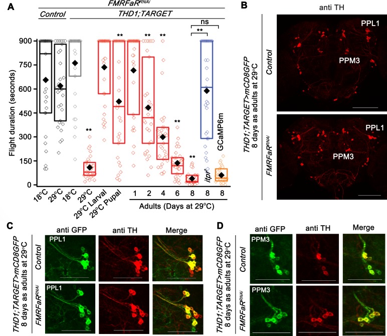
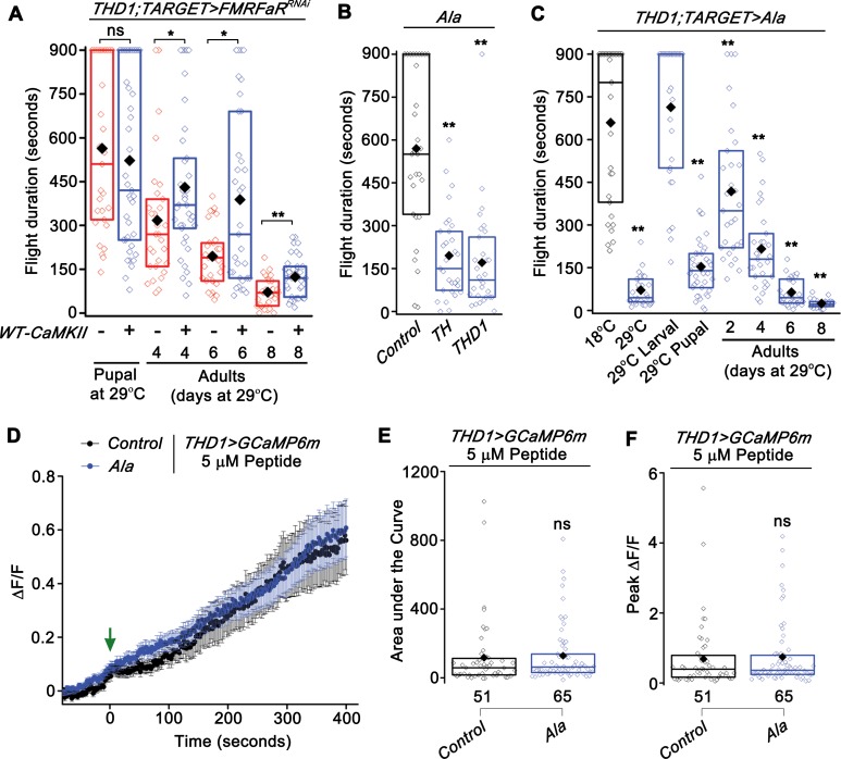
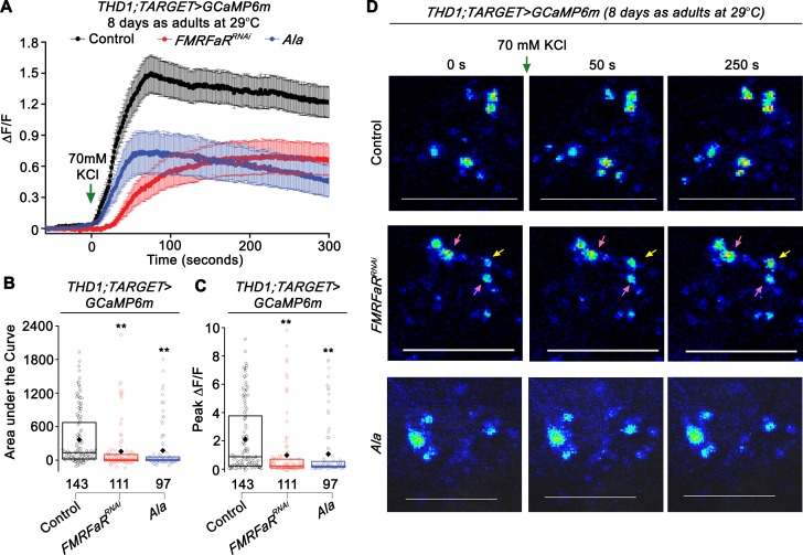
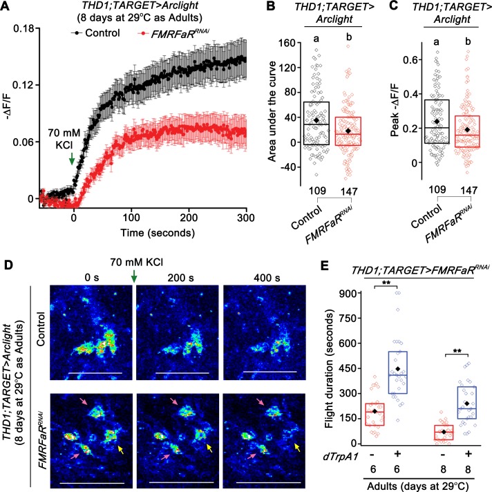
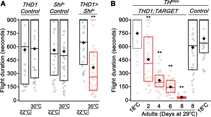
Neuropeptide signaling influences animal behavior by modulating neuronal activity and thus altering circuit dynamics. Insect flight is a key innate behavior that very likely requires robust neuromodulation. Cellular and molecular components that help modulate flight behavior are therefore of interest and require investigation. In a genetic RNAi screen for G-protein coupled receptors that regulate flight bout durations, we earlier identified several receptors, including the receptor for the neuropeptide FMRFa (FMRFaR). To further investigate modulation of insect flight by FMRFa we generated CRISPR-Cas9 mutants in the gene encoding the Drosophila FMRFaR. The mutants exhibit significant flight deficits with a focus in dopaminergic cells. Expression of a receptor specific RNAi in adult central dopaminergic neurons resulted in progressive loss of sustained flight. Further, genetic and cellular assays demonstrated that FMRFaR stimulates intracellular calcium signaling through the IP3R and helps maintain neuronal excitability in a subset of dopaminergic neurons for positive modulation of flight bout durations.


Conflict of interest statement

The authors have declared that no competing interests exist.

Figures

Fig 1
Fig 1. FMRFaR on dopaminergic neurons is required for flight.
(A) Box plot showing duration of flight bouts in flies expressing FMRFaRRNAi in the indicated neuronal domains (red). In every box plot, the limits extend from 25th to 75th percentile, the line and solid diamond represent the median and mean respectively and the open diamonds show the individual data points. Significance was calculated by comparing with either the control genotypes (FMRFaRRNAi/+, nSyb/+, TH/+, THD1/+) or as indicated by horizontal lines above the graph (n≥30, **p<0.01, Mann-Whitney U-test). Genotypic controls, referred to as ‘Control’ in the figure, have been obtained throughout by crossing the strains to be tested with wild-type, Canton-S. The ‘+’ in all control genotypes denotes the wild-type allele. (B) Quantitative PCR validation of FMRFaR knockdown in nSyb positive neurons. Each bar represents mean of normalized fold change ± SEM (n = 6, One-way ANOVA followed by post-hoc Tukey’s test; the same alphabet above each bar represents statistically indistinguishable groups; different alphabet represents p<0.01). (C) Snapshots of flight videos of the indicated genotypes (FMRFaRRNAi/+ and THD1>FMRFaRRNAi) at time points before and after air-puff stimulus (green arrow). Flies of the control genotype continued to fly for time periods beyond those shown in the images. (D) Representation of the FMRFaR gene locus. The CRISPR-Cas9 based deletion removes ~1500 bp of the 1650 bp coding sequence. Primers used to amplify the 5’ (pink: 5’F, 5’R) and 3’ (green: 3’F, 3’R) ends of the gene are indicated by arrows. Agarose gel showing PCR products from wild-type, heterozygous and homozygous knockouts of FMRFaR. L denotes DNA Ladder. (E) Quantitative PCR validation of FMRFaR CRISPR knockout from adult brains (n≥3, **p<0.01, unpaired t-test). (F) Flight bout duration of flies with CRISPR-Cas9 based knockout of FMRFaR. Statistical comparisons are as indicated by horizontal lines (n≥30, **p<0.01, Mann-Whitney U-test).
Fig 2
Fig 2. FMRFaR is expressed on dopaminergic neurons and is functionally active.
(A) Quantitative PCR on FACS sorted dopaminergic (GFP +ve, TH) and non-dopaminergic (GFP–ve, non TH) neurons, shows enrichment of FMRFaR transcripts in dopaminergic neurons. Each bar represents normalized fold change as mean ± SEM (n≥4, *p<0.05, unpaired t-test) (B) Snap shots of GCaMP6m responses from the indicated genotypes. The red box represents the magnified region of the protocerebrum shown in subsequent images. Warmer colors denote increase in [Ca2+]. Scale bars indicate 20 μm. (C, D) Mean traces (±SEM) of normalized GCaMP6m fluorescence responses (ΔF/F) in THD1GAL4 marked cells upon peptide or solvent addition (green arrow). (E) Area under the curve and (F) Peak ΔF/F quantified from 0 s to 420 s in (C, D). The box plot limits extend from 25th to 75th percentile. The line and solid diamond represent the median and mean respectively. Individual data points are shown as open diamonds. Numbers below indicate total number of cells imaged. Each bar is compared to the Control peptide stimulated condition shown in black (**p<0.01; ns—not significant; Mann-Whitney U-test).
Fig 3
Fig 3. FMRFaR is required in adult dopaminergic neurons for sustained flight.
(A) Box plot showing flight bout durations by temporal knockdown of FMRFaR in THD1 neurons using the TARGET system (THD1;TubGAL80ts>FMRFaRRNAi), which allows effective expression of transgene at 29°C, but not at 18°C. Animals maintained at 18°C or 29°C throughout development and as adults are indicated as 18°C and 29°C, respectively. Knockdown of FMRFaR only during the larval, pupal or adult stages are shown as 29°C Larval, 29°C Pupal or 29°C Adult, respectively (red). Knockdown of FMRFaR in adult stages (29°C Adult) reduced flight bout durations significantly. The phenotype observed upon FMRFaR knockdown in adults for 8 days could be rescued by overexpression of the itpr+ transgene, but not with another UAS transgene, GCaMP6m (THD1;TubGAL80ts>FMRFaRRNAi;itpr+ shown in blue and THD1;TubGAL80ts>FMRFaRRNAi;GCaMP6m shown in orange; n≥30, **p<0.01, ns–not significant; Mann-Whitney U-test). All comparisons are to the control condition, THD1;TARGET>FMRFaRRNAi at 18°C (grey bar; THD1;TubGAL80ts>FMRFaRRNAi; 18°C) or as indicated by horizontal lines above the graph (n≥30, **p<0.01, Mann-Whitney U-test). (B) Immunohistochemistry of representative adult brain samples corresponding to 8 day adult controls (above; THD1;TubGAL80ts>mCD8GFP Control) and FMRFaR knockdown (below; THD1;TubGAL80ts>mCD8GFP;FMRFaRRNAi). Anti-TH antibody was used to mark the various dopaminergic clusters, amongst which PPL1 and PPM3 clusters are indicated. Scale bars represent 100 μm. (C, D) Representative magnified views of the PPL1 (C) and PPM3 (D) clusters and their projections showing anti-GFP and anti-TH immunostaining in 8 day old adult brains of control (above; THD1;TubGAL80ts>mCD8GFP Control) and FMRFaR knockdown (below; THD1;TubGAL80ts>mCD8GFP;FMRFaRRNAi). Scale bar represents 50 μm.
Fig 4
Fig 4. CaMKII function is required in dopaminergic neurons to regulate flight.
(A) Flight bout durations observed with overexpression of WT-CaMKII in the background of FMRFaR knockdown in THD1 neurons (THD1;TubGAL80ts>WT-CaMKII;FMRFaRRNAi). A significant rescue of the flight defect was observed by expression in the adults, but not in pupae (n≥30, *p<0.05, **p<0.01, Mann-Whitney U-test). (B) Flight durations observed with expression of a genetically encoded CaMKII inhibitor peptide, Ala, in dopaminergic neurons as compared to Ala control (n≥30, **p<0.01, Mann-Whitney U-test). (C) Flight time of flies with temporal expression of Ala in THD1 neurons using the TARGET system (THD1;TubGAL80ts>Ala). Significant deficits in flight were observed upon CaMKII inhibition in pupal and adult stages as compared to flies maintained at 18°C throughout (n≥30, **p<0.01, Mann-Whitney U-test). (D) Mean traces (±SEM) of normalized GCaMP6m responses in control (THD1>GCaMP6m) and Ala expressing THD1 neurons (THD1>GCaMP6m;Ala) upon peptide stimulation (green arrow). (E) Area under the curve and (F) Peak ΔF/F quantified from 0 s to 420 s in (D) were not significantly different between the two genotypes tested (p>0.05; ns—not significant; Mann-Whitney U-test).
Fig 5
Fig 5. Expression of FMRFaRRNAi and inhibition of CaMKII in dopaminergic neurons reduces calcium entry after a depolarizing stimulus.
(A) Mean trace (±SEM) of normalized GCaMP6m responses observed upon KCl stimulation (green arrow) in 8 day adult neurons in the three indicated genotypes, THD1;TubGAL80ts>GCaMP6m, Control in black; THD1;TubGAL80ts>GCaMP6m;FMRFaRRNAi, in red and THD1;TubGAL80ts>GCaMP6m;Ala, in blue. (B) Area under the curve and (C) Peak change in fluorescence quantified from (A). Each bar is compared to control shown in black (**p<0.01; Mann-Whitney U-test). (D) Snap shots of GCaMP6m responses from the same genotypes shown in (A). In the FMRFaRRNAi knockdown condition (2nd row), the yellow arrow indicates a cell that responded whereas pink arrows indicate cells with no response or a minimal response to the depolarization stimulus. Scale bars represent 50 μm.
Fig 6
Fig 6. Expression of FMRFaRRNAi in dopaminergic neurons reduces membrane depolarization.
(A) Mean trace (±SEM) of negative change in fluorescence (-ΔF/F) observed upon KCl stimulation of THD1 neurons expressing a fluorescent-based membrane voltage indicator, Arclight in the indicated genotypes, THD1;TubGAL80ts>Arclight, Control in black; THD1;TubGAL80ts>Arclight;FMRFaRRNAi, in red. (B) Area under the curve and (C) Peak (-ΔF/F) quantified from (A) were significantly different in the FMRFaR knockdown neurons as compared to their genotypic controls. Numbers below each box plot indicate total number of cells imaged from a minimum of 5 independent brains (One-way ANOVA followed by post-hoc Tukey’s test; the same alphabet above each bar represents statistically indistinguishable groups; different alphabet represents p<0.05). (D) Representative images of neuronal depolarization as observed by a decrease in Arclight fluorescence in the same genotypes shown in (A). With FMRFaR knockdown (2nd row), few cells responded normally (yellow arrow), whereas most others showed no response or a minimal response to the depolarization stimulus (pink arrow). Scale bars represent 50 μm. (E) Rescue of flight bout durations by expression of dTrpA1 in the genetic background of FMRFaRRNAi in 6 and 8 day old adult THD1 marked neurons (THD1;TubGAL80ts>dTrpA1;FMRFaRRNAi compared to THD1;TubGAL80ts>FMRFaRRNAi; n≥30, **p<0.01, Mann-Whitney U-test).
Fig 7
Fig 7. Dopaminergic neurons require synaptic vesicle recycling for sustained flight.
(A) Acute expression of a temperature sensitive dynamin mutant (Shits) in adult dopaminergic neurons reduced flight bout durations when tested at 30°C as compared to a control tested at 22°C (THD1>Shits at 22°C compared to 30°C; n≥30, **p<0.01, Mann-Whitney U-test). THD1/+ and Shits/+ controls in the two temperature conditions are also shown. (B) Adult specific knockdown of the dopamine synthesizing enzyme, Tyrosine Hydroxylase (TH) by expressing an RNAi (THRNAi) in dopaminergic neurons significantly reduces flight duration as compared to the corresponding 18°C controls (THD1;TubGAL80ts>THRNAi; n≥30, **p<0.01, Mann-Whitney U-test).

Similar articles

Cited by

References

    1. van den Pol AN. Neuropeptide Transmission in Brain Circuits. Neuron. 2012;76: 98–115. 10.1016/j.neuron.2012.09.014 - DOI - PMC - PubMed
    1. Taghert PH, Nitabach MN. Peptide Neuromodulation in Invertebrate Model Systems. Neuron. 2012;76: 82–97. 10.1016/j.neuron.2012.08.035 - DOI - PMC - PubMed
    1. Griffith LC. Neuromodulatory control of sleep in Drosophila melanogaster: Integration of competing and complementary behaviors. Curr Opin Neurobiol. 2013;23: 819–823. 10.1016/j.conb.2013.05.003 - DOI - PMC - PubMed
    1. Kim SM, Su C, Wang JW. Neuromodulation of Innate Behaviors in Drosophila. Annu Rev Neurosci. 2017;40: 327–48. 10.1146/annurev-neuro-072116-031558 - DOI - PubMed
    1. Brembs B, Christiansen F, Pflüger HJ, Duch C, Pfluger HJ, Duch C. Flight Initiation and Maintenance Deficits in Flies with Genetically Altered Biogenic Amine Levels. J Neurosci. 2007;27: 11122–11131. 10.1523/JNEUROSCI.2704-07.2007 - DOI - PMC - PubMed

Publication types

MeSH terms

Substances