. 2018 Aug 15;18(1):819.
doi: 10.1186/s12885-018-4720-z.

Hypoxia promotes acquisition of aggressive phenotypes in human malignant mesothelioma

Affiliations

Hypoxia promotes acquisition of aggressive phenotypes in human malignant mesothelioma

Myung-Chul Kim et al. BMC Cancer. .

Abstract

Background: Hypoxia is a hallmark of the solid tumor microenvironment and is associated with poor outcomes in cancer patients. The present study was performed to investigate mechanisms underlying the hypoxia-induced phenotypic changes using human malignant mesothelioma (HMM) cells.

Methods: Hypoxic conditions were achieved by incubating HMM cells in the air chamber. The effect of hypoxia on phenotype changes in HMM cells was investigated by performing in vitro clonogenicity, drug resistance, migration, and invasion assays. Signaling pathways and molecules involved in the more aggressive behaviors of HMM cells under hypoxia were investigated. A two-tailed unpaired Student's t-test or one-way ANOVA with Bonferroni post-test correction was used in this study.

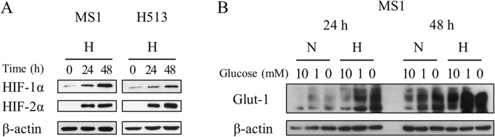
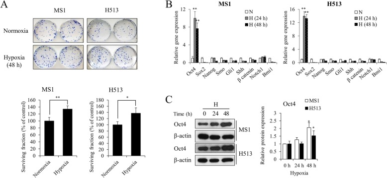
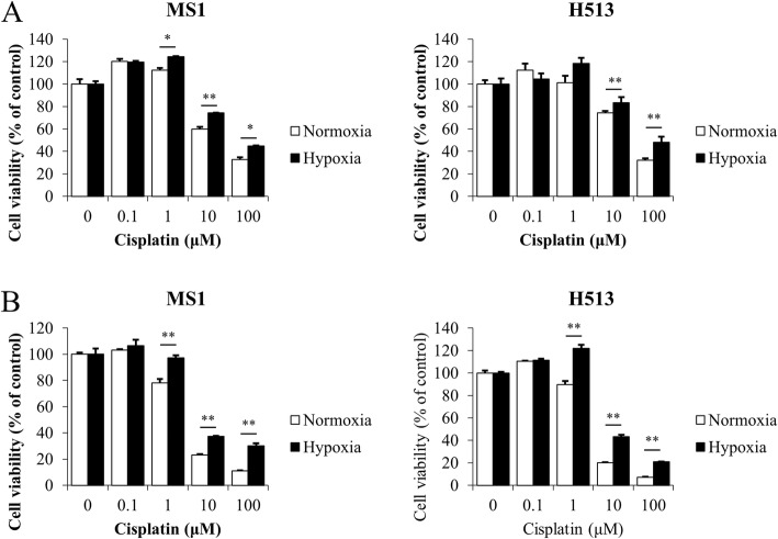
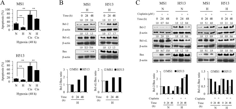
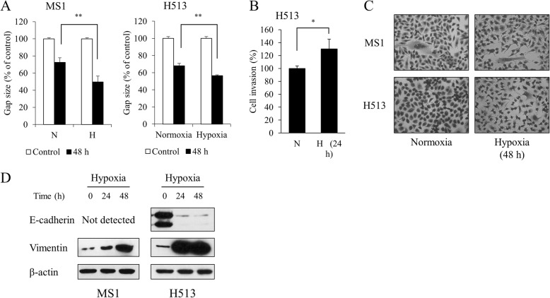
Results: Hypoxic conditions upregulated hypoxia-inducible factor 1 alpha (HIF-1α) and HIF-2α in parallel with the upregulation of its target, Glut-1, in HMM cells. In vitro clonogenicity of HMM cells was significantly increased in hypoxic conditions, but the proliferation of cells at a high density in hypoxia was lower than that in normoxic conditions. The expression levels of HIF-2α and Oct4 were increased in hypoxic HMM cells. The percentage of cells with high CD44 expression was significantly higher in HMM cells cultured in hypoxia than those cultured in normoxia. Hypoxia significantly enhanced the resistance of HMM cells to cisplatin, which occurred through cytoprotection against cisplatin-induced apoptosis. While cisplatin treatment decreased the ratio of Bcl-2 to Bax in normoxic condition, hypoxia conversely increased the ratio in HMM cells treated with cisplatin. Hypoxia increased the mobility and invasiveness of HMM cells. Epithelial to mesenchymal transition was promoted, which was indicated by the repression of E-cadherin and the concomitant increase of vimentin in HMM cells.

Conclusions: The data illustrated that hypoxic conditions augmented the aggressive phenotypes of HMM cells at the biological and molecular levels. The present study provides valuable background information beginning to understand aggressiveness of HMM in tumor microenvironments, suggesting that a control measure for tumor hypoxia may be an effective therapeutic strategy to reduce the aggressiveness of cancer cells in HMM patients.

Keywords: Drug resistance; EMT; HIFα; Hypoxia; Malignant phenotypes; Mesothelioma; Stemness; Tumor microenvironment.


Conflict of interest statement

Ethics approval and consent to participate

Not applicable.

Consent for publication

Not applicable.

Competing interests

The authors declare that they have no competing interests.

Publisher’s Note

Springer Nature remains neutral with regard to jurisdictional claims in published maps and institutional affiliations.

Figures

Fig. 1
Fig. 1
The experimental establishment of tumor hypoxia in HMM cells. (a) Hypoxia markedly increased HIF-1α expression and induced HIF-2α expression de novo in HMM cells. (b) A HIF-1/2α target Glut-1 increased in response to hypoxia and glucose starvation in MS1 cells. Abbreviations: N, normoxia; H, hypoxia
Fig. 2
Fig. 2
The effect of hypoxia on in vitro clonogenicity in HMM cells. (a) Hypoxia enhanced the colony forming ability of HMM cells. Representative microscopic examinations are presented. P value was calculated by Student’s t-test. Hypoxia significantly upregulated the expression of Oct4 at transcriptional (b) and translational (c) levels in HMM cells. P value was calculated by one-way ANOVA with Bonferroni post-test. *P value < 0.05, **P value < 0.01. Abbreviations: N, normoxia; H, hypoxia
Fig. 3
Fig. 3
The effect of hypoxia on cell proliferation in HMM cells. Hypoxia significantly decreased proliferation and viability in HMM cells at high cell seeding density. (a) Counting cell numbers. (b) MTT assay. The number of cells initially seeded is presented in parentheses. Cell cycle profiles did not appreciably differ between normoxic and hypoxic HMM cells (c). *P value < 0.05, **P value < 0.01, as calculated by Student’s t-test. Abbreviations: N, normoxia; H, hypoxia
Fig. 4
Fig. 4
The effect of hypoxia on drug sensitivity in HMM cells. MTT assay. The sensitivity to cisplatin was decreased in HMM cells under hypoxia for 24 h (a) and 48 h (b), compared to the sensitivity of those in normoxia. * indicates a significant difference compared with the corresponding normoxic cisplatin-treated control. *P value < 0.05, **P value < 0.01, as calculated by Student’s t-test
Fig. 5
Fig. 5
The effect of hypoxia on cisplatin-induced apoptosis in HMM cells. (a) Hypoxia significantly reduced apoptosis either with or without cisplatin in HMM cells. (b) Hypoxia upregulated the expression of anti-apoptotic Bcl-2, whereas Bcl-xL levels remained almost unchanged in HMM cells. Marked increase in pro-apoptotic Bax expression was detected in MS1 cells only. The ratio of Bcl-2 and Bcl-xL to Bax increased in H513 cells. (c) Hypoxia increased Bcl-2 levels and maintained the expression during exposure to cisplatin treatment in HMM cells, compared to the expression level in cells in normoxia. With cisplatin treatment, Bcl-xL expression either tended to decrease or remained unchanged in HMM cells under hypoxia, compared to the level in cells in normoxia. The cisplatin-induced increase in Bax expression was less under hypoxia than under normoxia in MS1 cells. The Bcl-2 to Bax ratio increased in HMM cells under hypoxia following cisplatin. All protein expression was normalized to endogenous β-actin level and is presented as densitometric values at the top of each protein blot. *P value < 0.05, as calculated by one-way ANOVA with Bonferroni post-test
Fig. 6
Fig. 6
The effect of hypoxia on migration and invasion in HMM cells. (a) Hypoxia significantly increased migration in HMM cells. (b) Hypoxia significantly enhanced invasion in H513 cells. (c) Phase contrast images (400× magnification) of HMM cells cultured for 48 h under normoxia or hypoxia are presented. (d) Hypoxia induced a loss of E-cadherin expression and a gain of vimentin expression in HMM cells. E-cadherin expression was not detected in MS1 cells. *P value < 0.05, as calculated by Student’s t-test

References

    1. Ruan K, Song G, Ouyang G. Role of hypoxia in the hallmarks of human cancer. J Cell Biochem. 2009;107(6):1053–1062. doi: 10.1002/jcb.22214. - DOI - PubMed
    1. Fukumura D, Jain RK. Tumor microenvironment abnormalities: causes, consequences, and strategies to normalize. J Cell Biochem. 2007;101(4):937–949. doi: 10.1002/jcb.21187. - DOI - PubMed
    1. Cosse J-P, Michiels C. Tumour hypoxia affects the responsiveness of cancer cells to chemotherapy and promotes cancer progression. Anti-Cancer Agent Me. 2008;8(7):790–797. doi: 10.2174/187152008785914798. - DOI - PubMed
    1. Vaupel P, Mayer A. Hypoxia in cancer: significance and impact on clinical outcome. Cancer Metast Rev. 2007;26(2):225–239. doi: 10.1007/s10555-007-9055-1. - DOI - PubMed
    1. Zhou J, Schmid T, Schnitzer S, Brüne B. Tumor hypoxia and cancer progression. Cancer Lett. 2006;237(1):10–21. doi: 10.1016/j.canlet.2005.05.028. - DOI - PubMed

MeSH terms

Substances