. 2018 Aug 15;17(1):122.
doi: 10.1186/s12943-018-0867-0.

Exosomes derived from mesenchymal stem cells enhance radiotherapy-induced cell death in tumor and metastatic tumor foci

Affiliations

Exosomes derived from mesenchymal stem cells enhance radiotherapy-induced cell death in tumor and metastatic tumor foci

Virgínea de Araujo Farias et al. Mol Cancer. .

Abstract

Background: We have recently shown that radiotherapy may not only be a successful local and regional treatment but, when combined with MSCs, may also be a novel systemic cancer therapy. This study aimed to investigate the role of exosomes derived from irradiated MSCs in the delay of tumor growth and metastasis after treatment with MSC + radiotherapy (RT).

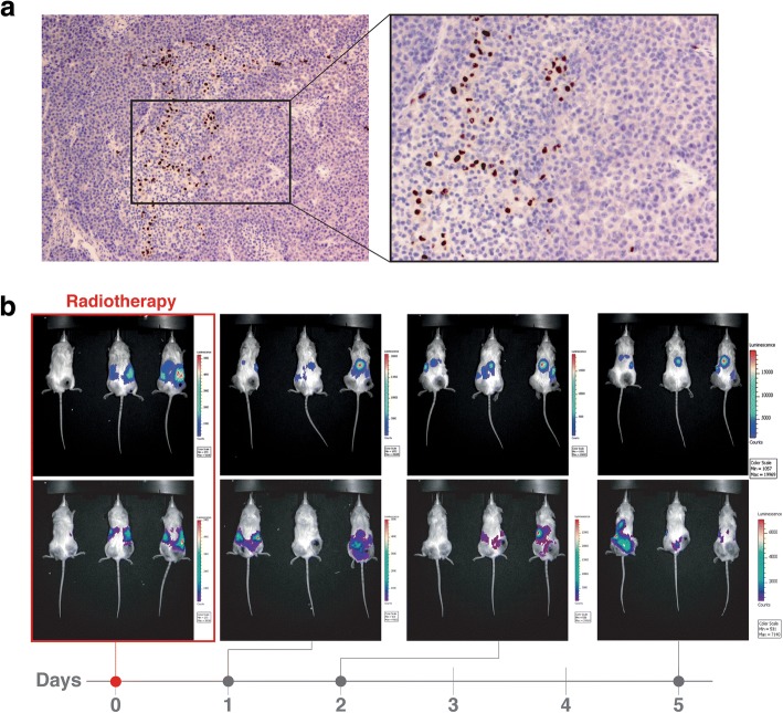
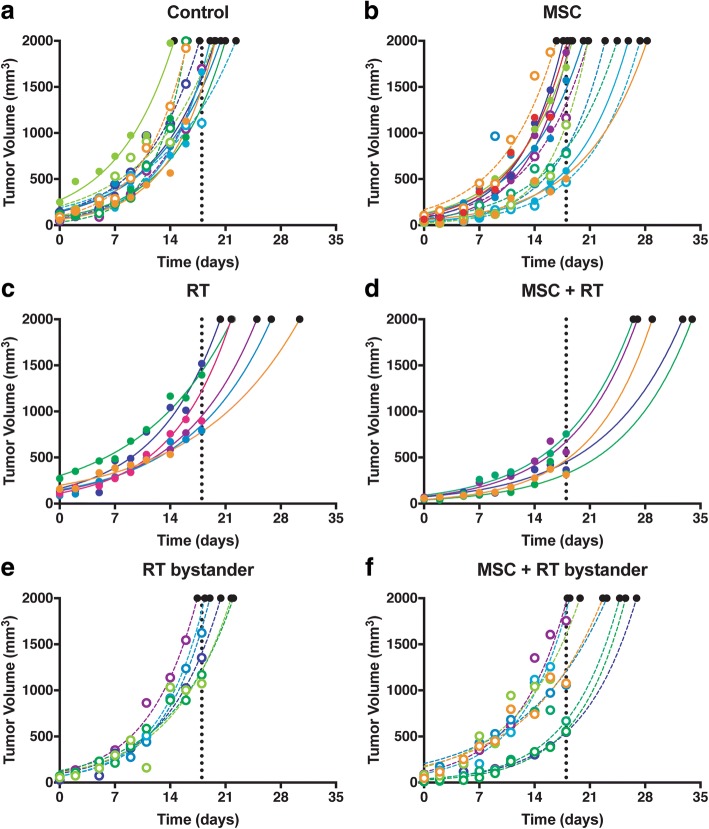
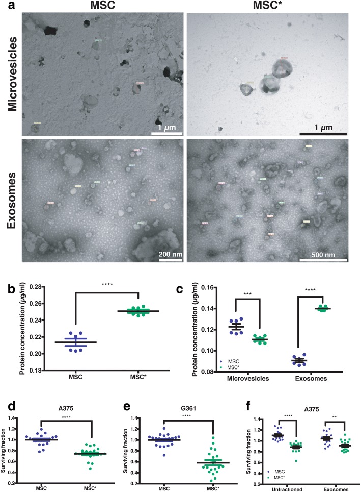
Methods: We have measured tumor growth and metastasis formation, of subcutaneous human melanoma A375 xenografts on NOD/SCID-gamma mice, and the response of tumors to treatment with radiotherapy (2 Gy), mesenchymal cells (MSC), mesenchymal cells plus radiotherapy, and without any treatment. Using proteomic analysis, we studied the cargo of the exosomes released by the MSC treated with 2 Gy, compared with the cargo of exosomes released by MSC without treatment.

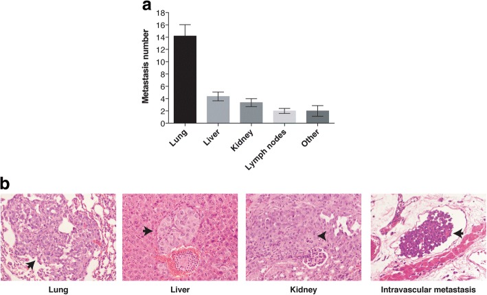
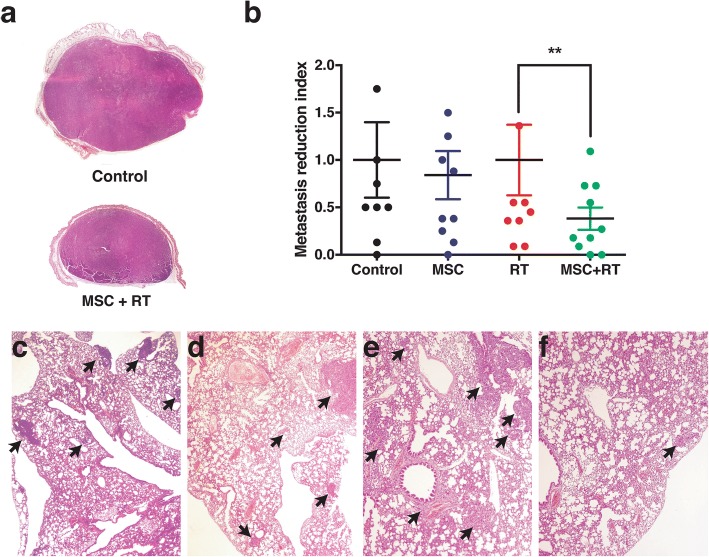
Results: The tumor cell loss rates found after treatment with the combination of MSC and RT and for exclusive RT, were: 44.4% % and 12,1%, respectively. Concomitant and adjuvant use of RT and MSC, increased the mice surviving time 22,5% in this group, with regard to the group of mice treated with exclusive RT and in a 45,3% respect control group. Moreover, the number of metastatic foci found in the internal organs of the mice treated with MSC + RT was 60% less than the mice group treated with RT alone. We reasoned that the exosome secreted by the MSC, could be implicated in tumor growth delay and metastasis control after treatment.

Conclusions: Our results show that exosomes derived form MSCs, combined with radiotherapy, are determinant in the enhancement of radiation effects observed in the control of metastatic spread of melanoma cells and suggest that exosome-derived factors could be involved in the bystander, and abscopal effects found after treatment of the tumors with RT plus MSC. Radiotherapy itself may not be systemic, although it might contribute to a systemic effect when used in combination with mesenchymal stem cells owing the ability of irradiated MSCs-derived exosomes to increase the control of tumor growth and metastasis.

Keywords: Abscopal effect; Annexin A1; Bystander effect; Cell therapy; Experimental radiotherapy; Melanoma xenograft; Mesenchymal stem cells; Metastasis spread; Proteomic analysis.


Conflict of interest statement

Ethics approval and consent to participate

These studies were performed in strict accordance with the recommendations of the Guide for the Care and Use of Laboratory Animals of the Bioethical Committee of Granada University, and the protocol was approved by the Committee on the Ethics of Animal Experiments of the CSIC.

Consent for publication

Not applicable.

Competing interests

The authors declare that they have no competing interests.

Publisher’s Note

Springer Nature remains neutral with regard to jurisdictional claims in published maps and institutional affiliations.

Figures

Fig. 1
Fig. 1
a Distribution of A375 xenografts’ micro-metastasis in the internal organs of the tumor-bearing NOD/SCID-gamma mice. Results are expressed as mean value ± standard error of mean. b Representative photomicrographs of H&E from lungs, liver, kidney and intravascular micro-metastasis (black arrow)
Fig. 2
Fig. 2
Biodistribution of MSC on tumor-bearing mice injected intratumorally or intraperitoneally. a Representative photomicrographs of A375 xenografts 24 after BrdU-labelled MSCs (brown) intratumoral injection. b Biodistribution of luciferase-labelled MSCs injected intraperitoneally on tumor-bearing NSG mice after receiving radiotherapy (right flank). The images were obtained at 0, 1, 2 and 5 days after injecting (intraperitoneally) 10 luciferase-expressing MSCs. Luciferin was injected 5 min before the images are obtained
Fig. 3
Fig. 3
Kinetics of individual tumor growth and its treatment response to (a) Control, (b) MSC, (c) RT, (d) MSC + RT, (e) bystander tumor after RT and (f) bystander tumor after MSC + RT. Black points at the of the extrapolated curves represents the time to tumor growth (T-t-G) values. The different colors on the graphs represent a different animal within each group. Full dots represent the treated tumor (when applicable) and open dots represent its contralateral tumor
Fig. 4
Fig. 4
a Mean values of the growth kinetic curves of the xenotumors infused on NSG mice. Different curves hat different slopes (P < 0,0001) and the differences between RT and RT + MSC curves are also significant (P < 0,0001). b Extrapolated values of the time to tumor growth until a volume of 2,00 ml. The treated tumors growth slowly than un-treated and bystander tumors. The mean value of tumors with RT + MSC is significantly longer than the T-t-G in the group treated with RT alone
Fig. 5
Fig. 5
Histopathological study of the internal organs of the tumor-bearing mice treated for only 15–17 days, (a) Representative panoramic images of A375 human melanoma xenografts from Control and MSC + RT. b Comparisons between the metastasis incidence of Control vs. MSC and RT vs. MSC + RT. MSC treatment alone had no effect on the metastasis incidence index. The combined treatment of MSC + RT diminished the early spread of metastases produced by the A375 xenograft. Representative photomicrographs of H&E lung micro-metastases from (c) Control, (d) MSC, (e) RT and (f) MSC + RT treatment groups
Fig. 6
Fig. 6
a Morphologic characterization of the extracellular vesicles released by MSC and MSC* precipitated by differential ultra-centrifugation. b Total protein concentration on the extracellular vesicles released by MSC and MSC*. c Protein concentration on the microvesicles and exosomes from MSC and MSC*. MSC or MSC* unfractioned conditioned medium reduced the surviving fractions of (d) A375 and (e) G361 cells. f Comparison between unfractioned conditioned medium from MSC and MSC* and of its exosomes on the A375 cell line. MSC conditioned medium (blue points) has been considered as the control as there is any statistical differences between MSC and growth media controls (data not shown). Differences are statistically significant between conditioned medium (P < 0.05) and exosomes (green points, P < 0.0001) from MSC and MSC*
Fig. 7
Fig. 7
Cluster analysis of the Gene Ontology (GO) Biological Process terms from the common and unique proteins identified in the exosomes of MSC and MSC*. Terms, represented as circles, are collapsed into clusters according to semantic similarities. The labels on the graphics represent the most unique terms. Circle colors reflect the p-values and circle sizes the number of identified proteins within the GO term. a MSC exosomesc. b Shared exosomes between MSC and irradiated MSC (MSC*). c MSC* exosomes

References

    1. Atun R, Jaffray DA, Barton MB, Bray F, Baumann M, Vikram B, Hanna TP, Knaul FM, Lievens Y, Lui TY, et al. Expanding global access to radiotherapy. The Lancet Oncology. 2015;16:1153–1186. doi: 10.1016/S1470-2045(15)00222-3. - DOI - PubMed
    1. Barton MB, Jacob S, Shafiq J, Wong K, Thompson SR, Hanna TP, Delaney GP. Estimating the demand for radiotherapy from the evidence: a review of changes from 2003 to 2012. Radiother Oncol. 2014;112:140–144. doi: 10.1016/j.radonc.2014.03.024. - DOI - PubMed
    1. Scaife JE, Barnett GC, Noble DJ, Jena R, Thomas SJ, West CM, Burnet NG. Exploiting biological and physical determinants of radiotherapy toxicity to individualize treatment. Br J Radiol. 2015;88:20150172. doi: 10.1259/bjr.20150172. - DOI - PMC - PubMed
    1. Burnet NG, Wurm R, Nyman J, Peacock JH: Normal tissue radiosensitivity--how important is it? Clinical oncology (Royal College of Radiologists (Great Britain)) 1996, 8:25–34. - PubMed
    1. Lopez E, Guerrero R, Nunez MI, del Moral R, Villalobos M, Martinez-Galan J, Valenzuela MT, Munoz-Gamez JA, Oliver FJ, Martin-Oliva D, Ruiz de Almodovar JM. early and late skin reactions to radiotherapy for breast cancer and their correlation with radiation-induced DNA damage in lymphocytes. Breast Cancer Res. 2005;7:R690–R698. doi: 10.1186/bcr1277. - DOI - PMC - PubMed

Publication types