

Mink (Neovison vison) kits with pre-weaning diarrhea have elevated serum amyloid A levels and intestinal pathomorphological similarities with New Neonatal Porcine Diarrhea Syndrome
- PMID: 30111375
- PMCID: PMC6094914
- DOI: 10.1186/s13028-018-0403-7
Mink (Neovison vison) kits with pre-weaning diarrhea have elevated serum amyloid A levels and intestinal pathomorphological similarities with New Neonatal Porcine Diarrhea Syndrome
Abstract
Background: Pre-weaning diarrhea (PWD) is a syndrome affecting farm-raised neonatal mink kits. Apart from diarrhea it causes greasy skin exudation, dehydration, and distressed behavior and can ultimately lead to death. No specific causative agents have been identified and the syndrome is regarded as multifactorial. The aim of the present study was to investigate a possible inflammatory state in mink kits with PWD, as indicated by raised serum concentrations of the acute phase protein serum amyloid A (SAA) and by changes in intestinal pathomorphology and intestinal contents of bacteria. Samples collected from 20 diarrheic mink kits with PWD and 20 age-matched non-diarrheic control mink kits from two commercial Danish farms during the pre-weaning period (April-May) in 2016 were analyzed.
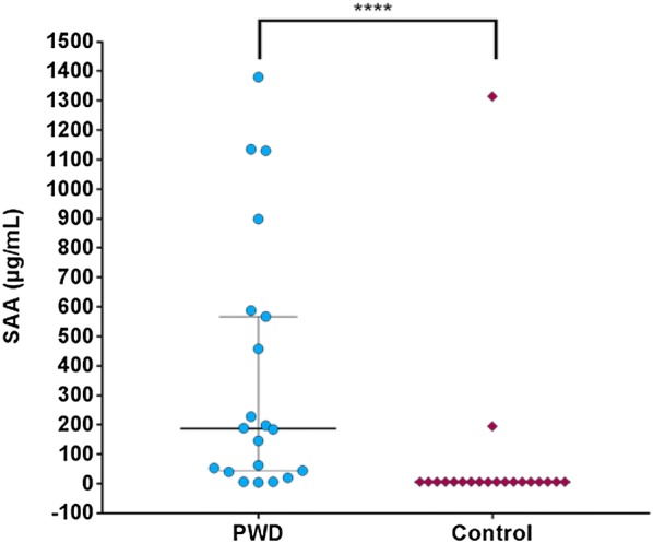
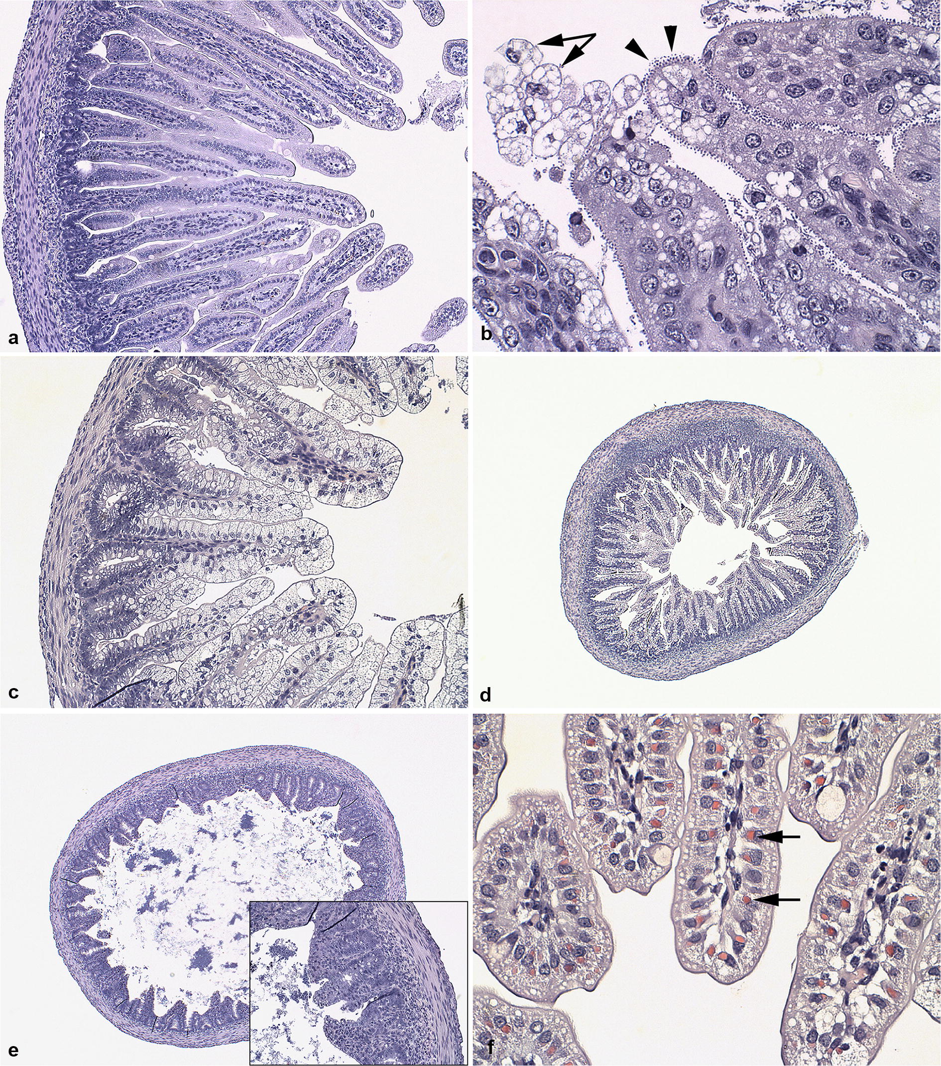
Results: Concentrations of SAA in serum samples from mink kits with PWD were significantly higher (up to 1000-fold) compared to non-diarrheic control mink kits. Significant features of enterocytic vacuolization, atrophy and fusion of villi in jejunum and mucosal atrophy of the colon of kits with PWD were found. Moreover, attachment of coccoid bacteria to enterocytes was more often found in kits suffering from PWD, while intra-cytoplasmic eosinophil bodies were more frequently observed in control kits. Cellular infiltrations with mononuclear and neutrophil leukocytes were not associated with disease status. Bacteria from the Staphylococcus intermedius group, such as Staphylococcus delphini, were more frequently cultivated from control mink kits, whereas Enterococcus spp. dominated in mink kits with PWD. Escherichia coli was cultivated from both control and mink kits with PWD, but with a higher frequency from mink kits with PWD.
Conclusion: A significant increase in circulating concentrations of SAA was found in PWD affected mink kits from 6 to 23 days old compared to controls. The histopathological changes in PWD mink kits suggest that the type of diarrhea is secretory. Attachment of coccoid bacteria, therefore, might be responsible for an enterotoxic effect causing a loss of balance in movements of ions and water leading to the vacuolization and swelling of the enterocytes. The slight to moderate infiltrations of neutrophils irrespectively of diarrheic status and the attachment of coccoid bacteria to enterocytes are comparable to observations found in piglets suffering from New Neonatal Porcine Diarrhea Syndrome. Mechanisms behind the correlation between increased SAA levels and the observed pathological intestinal features remain obscure.
Keywords: (Neovison vison); Bacteriology; Histology; Mink kits; Pre-weaning diarrhea; Serum amyloid A.
Figures
Similar articles
-
Low concentration of serum immunoglobulin G is associated with pre-weaning diarrhea in young mink kits (Neovison vison).Acta Vet Scand. 2019 Jun 10;61(1):26. doi: 10.1186/s13028-019-0461-5. Acta Vet Scand. 2019. PMID: 31182127 Free PMC article.
-
Investigation of the viral and bacterial microbiota in intestinal samples from mink (Neovison vison) with pre-weaning diarrhea syndrome using next generation sequencing.PLoS One. 2018 Oct 18;13(10):e0205890. doi: 10.1371/journal.pone.0205890. eCollection 2018. PLoS One. 2018. PMID: 30335814 Free PMC article.
-
Dam characteristics associated with pre-weaning diarrhea in mink (Neovison vison).Acta Vet Scand. 2018 Nov 12;60(1):73. doi: 10.1186/s13028-018-0427-z. Acta Vet Scand. 2018. PMID: 30419935 Free PMC article.
-
Post weaning diarrhea in pigs: risk factors and non-colistin-based control strategies.Acta Vet Scand. 2017 May 19;59(1):31. doi: 10.1186/s13028-017-0299-7. Acta Vet Scand. 2017. PMID: 28526080 Free PMC article. Review.
-
The Role of Probiotics in Alleviating Postweaning Diarrhea in Piglets From the Perspective of Intestinal Barriers.Front Cell Infect Microbiol. 2022 May 30;12:883107. doi: 10.3389/fcimb.2022.883107. eCollection 2022. Front Cell Infect Microbiol. 2022. PMID: 35711653 Free PMC article. Review.
Cited by
-
Detection of mink astrovirus in Poland and further phylogenetic comparison with other European and Canadian astroviruses.Virus Genes. 2021 Jun;57(3):258-265. doi: 10.1007/s11262-021-01834-z. Epub 2021 Apr 15. Virus Genes. 2021. PMID: 33860418 Free PMC article.
References
-
- Chriél M. Greasy kits-an efficient effort limits losses. Dansk Pelsdyravl. 1994;4:180–181.
-
- Svennekjær NC. Mink diseases and current problems in mink farming. Dansk Pelsdyravl. 1954;12:384–404.
-
- Hyldgaard-Jensen C. “Greasy kits” in mink. Observations over a three year period. Dansk Veterinærtidskrift. 1989;72:566–571.
-
- Clausen TN, Dietz HH. Greasy kits. Annual Report, Kopenhagen Research. Kopenhagen Fur. 2004; p. 209–14 (in Danish).
MeSH terms
Substances
LinkOut - more resources
Full Text Sources
Other Literature Sources
Medical
Miscellaneous
