

Detection of GNAS mutations in intramuscular / cellular myxomas as diagnostic tool in the classification of myxoid soft tissue tumors
- PMID: 30111377
- PMCID: PMC6094570
- DOI: 10.1186/s13000-018-0734-8
Detection of GNAS mutations in intramuscular / cellular myxomas as diagnostic tool in the classification of myxoid soft tissue tumors
Abstract
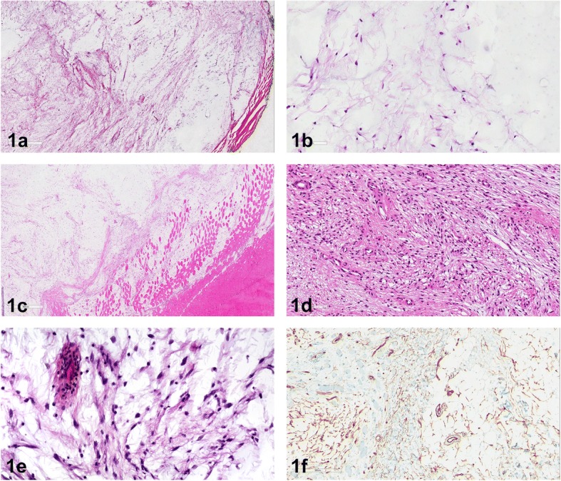
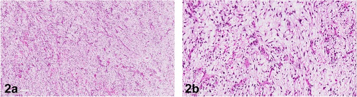
Background: Intramuscular / cellular myxomas and low-grade myxofibrosarcomas are two different tumor entities with a significant histological overlap, especially if dealing with small biopsies. Despite the morphological similarities, they differ considerably in their biological behaviour. Intramuscular / cellular myxoma rarely shows signs of recurrence and never metastasizes, in contrast to myxofibrosarcoma that tends to recur more aggressively and to metastasize haematologically. Therefore, it is of great importance to distinguish these lesions - evaluation of GNAS mutation status could be of tremendous help.
Methods: We reviewed 13 cases with intramuscular / cellular myxomas. The 13 cases included 5 men and 8 women, aged from 33 to 71 years (mean age 55.5 years). Immunohistochemistry was performed as well as next generation sequencing. Ten cases were located in the lower extremities and three cases were located in the upper extremities. Two lesions were initially misdiagnosed as a low-grade myxofibrosarcoma.
Results: Performing next generation sequencing 12 out of 13 specimens showed a GNAS mutation.
Conclusions: Our findings demonstrate that GNAS mutations are more common in intramuscular / cellular myxomas, than had been reported in literature in the past. Next generation sequencing for determining GNAS mutation status on small biopsies or diagnostically challenging cases facilitates the diagnosis of intramuscular / cellular myxoma and separates this tumor entity from its mimics.
Keywords: GNAS mutation; Intramuscular / cellular myxoma; Myxofibrosarcoma; Soft tissue pathology.
Conflict of interest statement
Ethics approval and consent to participate
The submitted study was part of a large study on myxofibrosarcomas. All patients and patients’ data were anonymized. The study was performed according to the institutional guidelines and approved by the Ethics committee of the Medical University of Graz (Vote 26–524 ex13/14). Patients were treated at the Department of Orthopaedics and Trauma, Medical University of Graz. The patients signed a general institutional consent, that their tissue and data can be used for research and publications.
Consent for publication
Not applicable.
Competing interests
The authors declare that they have no competing interests.
Publisher’s Note
Springer Nature remains neutral with regard to jurisdictional claims in published maps and institutional affiliations.
Figures
Similar articles
-
The Role of Methylation Analysis in Distinguishing Cellular Myxoma from Low-Grade Myxofibrosarcoma.Int J Mol Sci. 2024 May 8;25(10):5105. doi: 10.3390/ijms25105105. Int J Mol Sci. 2024. PMID: 38791144 Free PMC article.
-
Identification of novel GNAS mutations in intramuscular myxoma using next-generation sequencing with single-molecule tagged molecular inversion probes.Diagn Pathol. 2019 Feb 8;14(1):15. doi: 10.1186/s13000-019-0787-3. Diagn Pathol. 2019. PMID: 30736805 Free PMC article.
-
Karyotyping and analysis of GNAS locus in intramuscular myxomas.Oncotarget. 2017 Mar 28;8(13):22086-22094. doi: 10.18632/oncotarget.14986. Oncotarget. 2017. PMID: 28160572 Free PMC article.
-
Novel Association of Odontogenic Myxoma with Constitutional Chromosomal 1q21 Microduplication: Case Report and Review of the Literature.Pediatr Dev Pathol. 2016 Mar-Apr;19(2):139-45. doi: 10.2350/15-05-1637-CR.1. Epub 2015 Jul 31. Pediatr Dev Pathol. 2016. PMID: 26230961 Review.
-
Myxoma is not a single entity: a review of the concept of myxoma.Ann Diagn Pathol. 2000 Apr;4(2):99-123. doi: 10.1016/s1092-9134(00)90019-4. Ann Diagn Pathol. 2000. PMID: 10760324 Review.
Cited by
-
Artificial intelligence significantly improves the diagnostic accuracy of deep myxoid soft tissue lesions in histology.Sci Rep. 2022 Apr 28;12(1):6965. doi: 10.1038/s41598-022-11009-x. Sci Rep. 2022. PMID: 35484289 Free PMC article.
-
Surgical Treatment of Intramuscular Myxoma.Indian J Orthop. 2021 Feb 22;55(4):892-897. doi: 10.1007/s43465-021-00367-9. eCollection 2021 Aug. Indian J Orthop. 2021. PMID: 34194644 Free PMC article.
-
The Role of Methylation Analysis in Distinguishing Cellular Myxoma from Low-Grade Myxofibrosarcoma.Int J Mol Sci. 2024 May 8;25(10):5105. doi: 10.3390/ijms25105105. Int J Mol Sci. 2024. PMID: 38791144 Free PMC article.
-
Mazabraud's syndrome in female patients: Two case reports.World J Orthop. 2024 Jun 18;15(6):593-601. doi: 10.5312/wjo.v15.i6.593. eCollection 2024 Jun 18. World J Orthop. 2024. PMID: 38947265 Free PMC article.
-
Intramuscular myxoma of the biceps brachii muscle: a case report.J Surg Case Rep. 2022 Apr 11;2022(4):rjac145. doi: 10.1093/jscr/rjac145. eCollection 2022 Apr. J Surg Case Rep. 2022. PMID: 35422994 Free PMC article.
References
-
- Fletcher CD, Hogendoorn P, Mertens F, Bridge J. WHO classification of tumours of soft tissue and bone. 4th ed. Lyon: IARC Press; 2013.
-
- Hornick JL. Practical soft tissue pathology: a diagnostic approach. 1st ed. Philadelphia: Elsevier Saunders; 2013.
MeSH terms
Substances
Grants and funding
LinkOut - more resources
Full Text Sources
Other Literature Sources
Miscellaneous
