

Dropping in on lipid droplets: insights into cellular stress and cancer
- PMID: 30111611
- PMCID: PMC6146295
- DOI: 10.1042/BSR20180764
Dropping in on lipid droplets: insights into cellular stress and cancer
Abstract
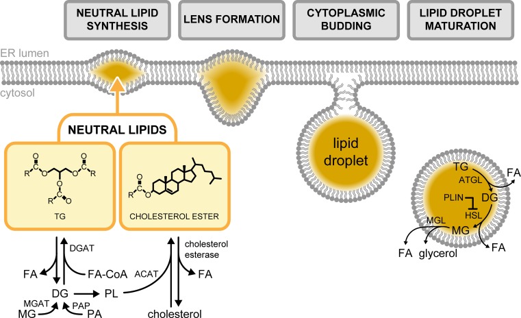
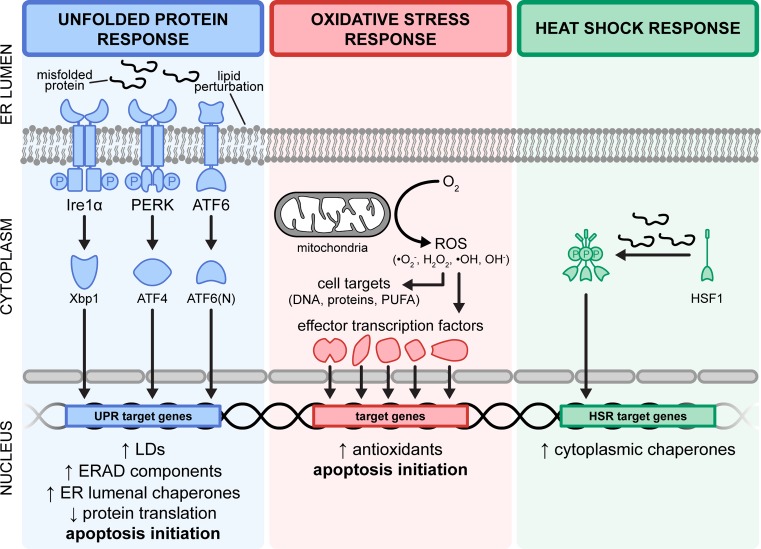
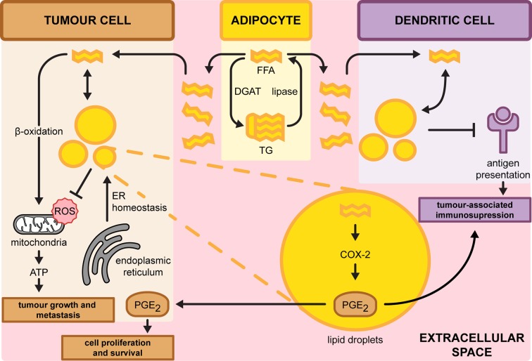
Lipid droplets (LD) have increasingly become a major topic of research in recent years following its establishment as a highly dynamic organelle. Contrary to the initial view of LDs being passive cytoplasmic structures for lipid storage, studies have provided support on how they act in concert with different organelles to exert functions in various cellular processes. Although lipid dysregulation resulting from aberrant LD homeostasis has been well characterised, how this translates and contributes to cancer progression is poorly understood. This review summarises the different paradigms on how LDs function in the regulation of cellular stress as a contributing factor to cancer progression. Mechanisms employed by a broad range of cancer cell types in differentially utilising LDs for tumourigenesis will also be highlighted. Finally, we discuss the potential of targeting LDs in the context of cancer therapeutics.
Keywords: cancer progression; cell stress; chemoresistance; lipid droplets.
© 2018 The Author(s).
Conflict of interest statement
The authors declare that there are no competing interests associated with the manuscript.
Figures
Similar articles
-
Friend or Foe: Lipid Droplets as Organelles for Protein and Lipid Storage in Cellular Stress Response, Aging and Disease.Molecules. 2020 Oct 30;25(21):5053. doi: 10.3390/molecules25215053. Molecules. 2020. PMID: 33143278 Free PMC article. Review.
-
Models of lipid droplets growth and fission in adipocyte cells.Exp Cell Res. 2015 Aug 15;336(2):253-62. doi: 10.1016/j.yexcr.2015.06.001. Epub 2015 Jun 26. Exp Cell Res. 2015. PMID: 26121906
-
Rab proteins as regulators of lipid droplet formation and lipolysis.Cell Biol Int. 2016 Oct;40(10):1026-32. doi: 10.1002/cbin.10650. Epub 2016 Sep 5. Cell Biol Int. 2016. PMID: 27453349 Review.
-
Lipid Droplets Big and Small: Basic Mechanisms That Make Them All.Annu Rev Cell Dev Biol. 2024 Oct;40(1):143-168. doi: 10.1146/annurev-cellbio-012624-031419. Annu Rev Cell Dev Biol. 2024. PMID: 39356808 Review.
-
Quantitative high-content/high-throughput microscopy analysis of lipid droplets in subject-specific adipogenesis models.Cytometry A. 2017 Nov;91(11):1068-1077. doi: 10.1002/cyto.a.23265. Epub 2017 Oct 14. Cytometry A. 2017. PMID: 29031005
Cited by
-
Oxidative stress mediates the inhibitory effects of Manzamine A on uterine leiomyoma cell proliferation and extracellular matrix deposition via SOAT inhibition.Redox Biol. 2023 Oct;66:102861. doi: 10.1016/j.redox.2023.102861. Epub 2023 Aug 25. Redox Biol. 2023. PMID: 37666118 Free PMC article.
-
Metabolic Plasticity in Chemotherapy Resistance.Front Oncol. 2020 Mar 6;10:281. doi: 10.3389/fonc.2020.00281. eCollection 2020. Front Oncol. 2020. PMID: 32211323 Free PMC article. Review.
-
Exploiting directed self-assembly and disassembly for off-to-on fluorescence responsive live cell imaging.RSC Adv. 2022 Dec 13;12(55):35655-35665. doi: 10.1039/d2ra06534g. eCollection 2022 Dec 12. RSC Adv. 2022. PMID: 36545082 Free PMC article. Review.
-
Multiplex Stimulated Raman Scattering Imaging Cytometry Reveals Lipid-Rich Protrusions in Cancer Cells under Stress Condition.iScience. 2020 Mar 27;23(3):100953. doi: 10.1016/j.isci.2020.100953. Epub 2020 Feb 29. iScience. 2020. PMID: 32179477 Free PMC article.
-
Novel CF₃-Substituted Pyridine- and Pyrimidine-Based Fluorescent Probes for Lipid Droplet Bioimaging.Int J Mol Sci. 2025 May 30;26(11):5271. doi: 10.3390/ijms26115271. Int J Mol Sci. 2025. PMID: 40508080 Free PMC article.
References
-
- Fawcett D.W. (1955) Observations on the cytology and electron microscopy of hepatic cells. J. Natl. Cancer Inst. 15, 1475–1503 - PubMed
Publication types
MeSH terms
Substances
LinkOut - more resources
Full Text Sources
Other Literature Sources
Research Materials