. 2018 Aug 15;10(454):eaap9527.
doi: 10.1126/scitranslmed.aap9527.

A protective Langerhans cell-keratinocyte axis that is dysfunctional in photosensitivity

Affiliations

A protective Langerhans cell-keratinocyte axis that is dysfunctional in photosensitivity

William D Shipman et al. Sci Transl Med. .

Abstract

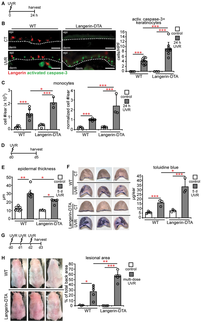
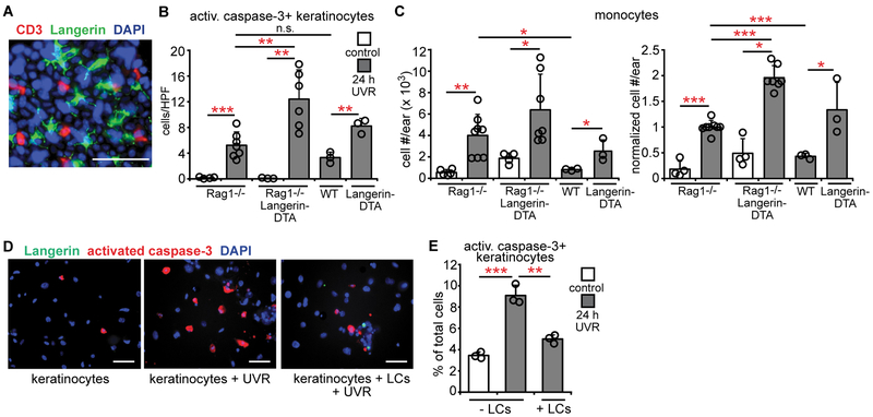
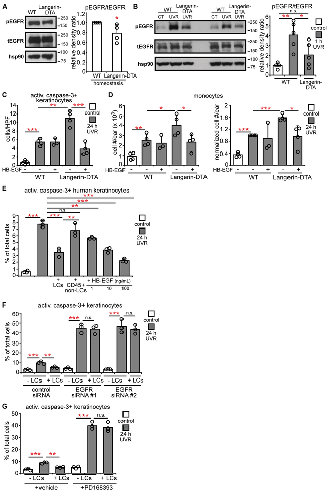
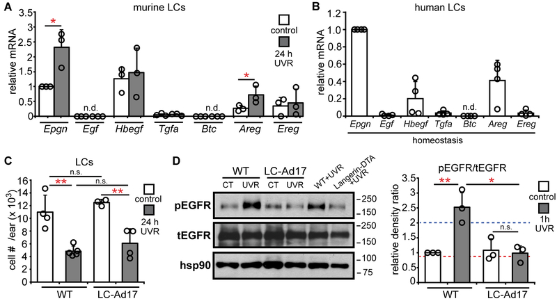
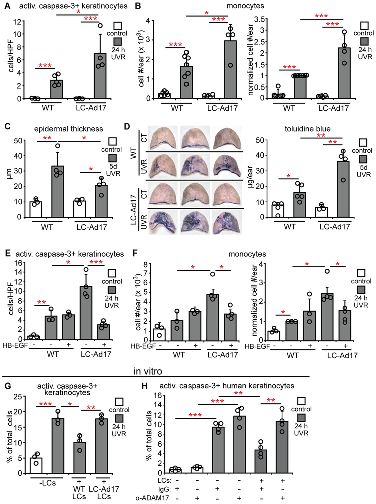
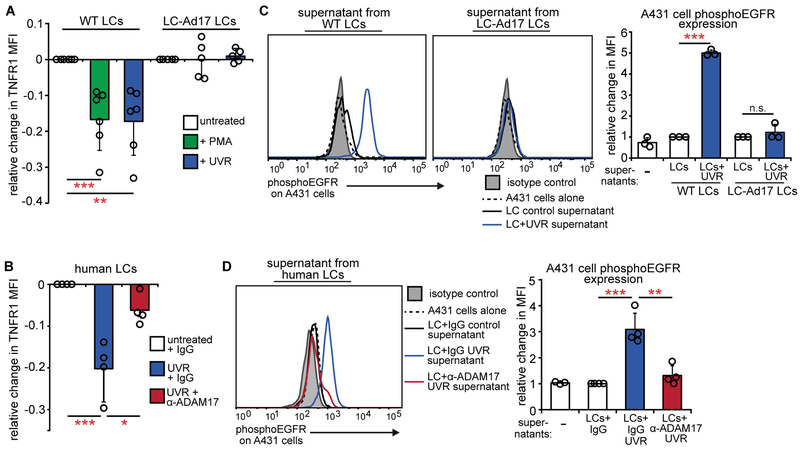
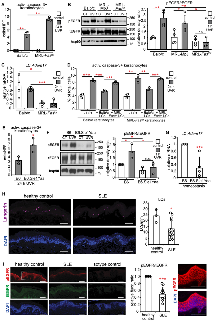
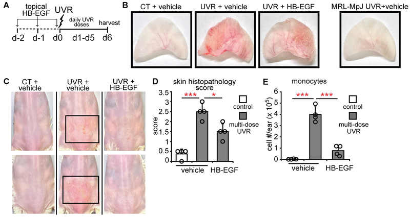
Photosensitivity, or skin sensitivity to ultraviolet radiation (UVR), is a feature of lupus erythematosus and other autoimmune and dermatologic conditions, but the mechanistic underpinnings are poorly understood. We identify a Langerhans cell (LC)-keratinocyte axis that limits UVR-induced keratinocyte apoptosis and skin injury via keratinocyte epidermal growth factor receptor (EGFR) stimulation. We show that the absence of LCs in Langerin-diphtheria toxin subunit A (DTA) mice leads to photosensitivity and that, in vitro, mouse and human LCs can directly protect keratinocytes from UVR-induced apoptosis. LCs express EGFR ligands and a disintegrin and metalloprotease 17 (ADAM17), the metalloprotease that activates EGFR ligands. Deletion of ADAM17 from LCs leads to photosensitivity, and UVR induces LC ADAM17 activation and generation of soluble active EGFR ligands, suggesting that LCs protect by providing activated EGFR ligands to keratinocytes. Photosensitive systemic lupus erythematosus (SLE) models and human SLE skin show reduced epidermal EGFR phosphorylation and LC defects, and a topical EGFR ligand reduces photosensitivity. Together, our data establish a direct tissue-protective function for LCs, reveal a mechanistic basis for photosensitivity, and suggest EGFR stimulation as a treatment for photosensitivity in lupus erythematosus and potentially other autoimmune and dermatologic conditions.


Figures

Fig. 1.
Fig. 1.. LCs limit UVR-induced keratinocyte apoptosis and skin injury.
(A-H) WT and Langerin-DTA mice were exposed to UVR and examined. (A) Experimental scheme for (B,C); ears were harvested 24 hours after UVR. (B) Activated caspase-3+ keratinocytes per high powered field (HPF). Left: Representative images of Langerin (red) and activated caspase-3 (green) stain. Right: Quantification (n= 3-9 mice). Scale bars: 50 μm. (C) Absolute (left) and normalized (right) monocyte numbers assessed by flow cytometry (n= 3-7). (D) Experimental scheme for (E,F); ears were harvested 5 days after UVR. (E) Epidermal thickness (n= 3-7 mice). (F) Epidermal permeability as assessed by toluidine blue penetrance. Left: Representative images. Right: Quantification (n= 3-5 mice). (G) Experimental scheme for (H); mice were exposed to UVR for 3 days and examined 24 hours later. (H) Left: Representative images of back skin. Right: Lesional area quantification (n= 3-5 mice). Bars represent means. (B,C,F,H) or medians (E). Error bars depict standard deviations (B,C,F,H) or interquartile ranges (E). *p<0.05, **p<0.01, ***p<0.001 using two-tailed unpaired Student’s t-test (B,C,F,H) or nonparametric non-directional Mann-Whitney U test (E) after one-way analysis of variance (ANOVA). Data are from 9 (B), 5 (C), 4 (E), 2 (F), and 3 (H) independent experiments.
Fig. 2.
Fig. 2.. LCs limit UVR-induced keratinocyte apoptosis directly.
(A) Whole mount stain of homeostatic mouse epidermis for CD3 (red), Langerin (green), and DAPI (blue). (B,C) Rag1−/−, Rag1−/−Langerin-DTA, WT, and Langerin-DTA mice were exposed to UVR and ears were harvested 24 hours later (n= 3-8 mice). (B) Activated caspase-3+ keratinocytes. (C) Absolute (left) and normalized (right) monocyte numbers. (D,E) Effect of LCs on keratinocyte survival in vitro. Murine keratinocyte cultures without and with LCs were exposed to UVR and examined 24 hours later (n= 3 mice). (D) Representative images of cultures stained for Langerin (green), activated caspase-3 (red), and DAPI (blue). (E) Activated caspase-3+ keratinocytes. (E) Data are from 5 (B,C) and 3 (A,D,E) independent experiments. Scale bars: 50 μm. (B,C,E) Bars represent means. Error bars depict standard deviations. *p<0.05, **p<0.01, ***p<0.001 using two-tailed unpaired Student’s t-test after one-way ANOVA.
Fig. 3.
Fig. 3.. LCs are required for UVR-induced epidermal EGFR activation and protect keratinocytes via EGFR stimulation.
(A,B) Epidermal EGFR phosphorylation at homeostasis (A) and 1 hour after UVR (B) (n= 4-5 mice). Left: Representative Western blot for phosphoEGFR (pEGFR), total EGFR (tEGFR), and hsp90 (loading control). Right: pEGFR:tEGFR relative density ratio. Uncropped blots in Fig. S13. (C,D) Mouse ears were treated with vehicle or HB-EGF prior to UVR and examined 24 hours after UVR (n= 3-4 mice). (C) Activated caspase-3+ keratinocytes. (D) Absolute (left) and normalized (right) monocyte numbers. (E) Effect of human LCs on UVR-induced keratinocyte apoptosis. Primary human keratinocytes without or with indicated cells or recombinant HB-EGF were exposed to UVR and examined 24 hours later (n= 3 human donors). (F,G) Effect of keratinocyte EGFR knockdown and inhibition on LC-mediated protection. Primary murine keratinocytes were treated with EGFR-targeted or control siRNAs (F) or PD168393 (G) before LC co-culture and UVR exposure (n= 3 mice). Bars represent means. Error bars depict standard deviations. Data are from 2 (A,B,F,G), 4 (C,D), and 3 (E) independent experiments. *p< 0.05, **p< 0.01, ***p< 0.001 using two-tailed unpaired Student’s t-test. T-test was performed after one-way ANOVA for (B-G).
Fig. 4.
Fig. 4.. LCs express EGFR ligands and LC-derived ADAM 17 mediates UVR-induced epidermal EGFR phosphorylation.
(A,B) Murine (A) and human (B) LC EGFR ligand expression (n=3-4 mice or human donors). Murine LCs were sorted from control or UVR-exposed mice. Expression of each ligand was normalized to control murine Epgn or human Epgn expression. (C,D) WT and LC-Ad17 mice were treated with UVR and analyzed at indicated time points. (C) LC numbers (n= 3-5 mice). (D) Epidermal EGFR phosphorylation. Left: Representative Western blot. Right: pEGFR:tEGFR ratio. Dashed lines are the values for the UVR-exposed WT (blue) and Langerin-DTA (red) mice shown in the blot. Uncropped blots in Fig. S13. Data are from 3 (A,B), 4 (C), and 2 (D) independent experiments. Bars represent means. Error bars depict standard deviations. n.s.= not significant p≥0.05, *p<0.05, **p<0.01 using two-tailed unpaired Student’s t-test. T-test was performed after one-way ANOVA for (C,D).
Fig. 5.
Fig. 5.. LC-derived ADAM 17 limits UVR-induced keratinocyte apoptosis and skin injury.
(A-D) WT and LC-Ad17 mice were treated with UVR and analyzed at indicated time points. (A) Activated caspase-3+ keratinocytes (n= 3-5 mice). (B) Absolute (left) and normalized (right) monocyte numbers (n= 4-7 mice). (C) Epidermal thickness (n= 3-mice). (D) Epidermal permeability (n= 3-5 mice). Left: Representative images. Right: Quantification. (E,F) Vehicle or HB-EGF was applied on the ears prior to UVR exposure (n= 3-4 mice). (E) Activated caspase-3+ keratinocytes. (F) Absolute (left) and normalized (right) monocyte numbers. (G,H) Effect of LC Adam17 deletion or ADAM17 blockade on keratinocyte survival in vitro. Murine keratinocytes with LCs from indicated mice (G) and human keratinocytes with control-IgG or anti-ADAM17-treated LCs (H) were exposed to UVR and examined at 24 hours (n= 3 mice or 4 human donors). Data are from 3 (E-G), 4 (A), 2 (H), 5 (B), and 1 (C,D) independent experiments. Bars represent means (A,B,D-H) or medians (C). Error bars depict standard deviations (A,B,D-H) or interquartile ranges (C). n.s.= not significant p≥0.05, *p<0.05, **p<0.01, ***p<0.001 using two-tailed unpaired Student’s t-test (A,B,D-H) or nonparametric non-directional Mann-Whitney U test (C) after one-way ANOVA.
Fig. 6.
Fig. 6.. UVR directly activates LC ADAM17 and EGFR ligand release.
(A, B) Effect of UVR on ADAM17 activity in sorted murine (A) and human (B) LCs as measured by change in TNFR1 mean fluorescence intensity (MFI) 45 minutes after the indicated treatments. PMA is a positive control. (n= 5-6 mice; n=4 human donors). (C,D) Conditioned supernatants from murine (C) or human LCs (D) were added to A431 EGFR indicator cells and phosphoEGFR was measured 10 minutes later by flow cytometry. Murine LC supernatants were from (A); human LC supernatants were from cells treated similarly to (B), except that antibody was washed out prior to UVR (see Supplementary Methods). Left: Representative histogram. Right: Quantification relative to cells treated with control WT LC supernatants (C) or control IgG-treated LC supernatants (D). Results are from 6 (A), 2 (B,D), and 3 (C) independent experiments. Bars represent means. Error bars depict standard deviations. n.s.= not significant p≥0.05, *p<0.05, **p<0.01, ***p<0.001 using two-tailed unpaired Student’s t-test after one-way ANOVA.
Fig. 7.
Fig. 7.. Photosensitive SLE mouse models and human SLE skin show a dysfunctional LC-keratinocyte axis.
WT and MRL-Faslpr(n= 2-4 mice) (A-C) or B6.Sle1Yaa mice (n= 3-5 mice) (E-G) were treated and examined as indicated. (A,E) Activated caspase-3+ keratinocytes. (B,F) Epidermal EGFR phosphorylation 1 hour after UVR. Left: Representative Western blot. Right: pEGFR:tEGFR ratio. Uncropped Western blots in Fig. S13. (C,G) LC Adam17 expression. (D) Effect of MRL-Faslpr LCs on keratinocyte apoptosis. Balb/c or MRL-Faslpr keratinocytes were exposed to UVR without or with indicated LCs. (n= 3 mice). (H,I) LC numbers and epidermal EGFR phosphorylation in human SLE skin (n= 3 healthy controls, 10-13 SLE patients). (H) Left: Representative images of anti-Langerin (purple) and DAPI (blue) staining. Right: LC numbers per mm of tissue. (I) Left: Representative images of anti-pEGFR (red), anti-tEGFR (green), and DAPI (blue) staining. Middle: Relative pEGFR:tEGFR fluorescence intensity normalized to healthy control skin. Right: Magnified inset from pEGFR and DAPI stain. Data are from 3 (A,B,D,E,G-I), and 2 (C,F) independent experiments. Bars represent means. Error bars depict standard deviations. n.s= not significant p≥0.05, *p<0.05, **p<0.01, ***p<0.001 using two-tailed unpaired Student’s t-test. T-test was performed after one-way ANOVA for (A-D, F).
Fig. 8.
Fig. 8.. Topical EGFR ligand reduces photosensitivity.
(A) Experimental scheme for (B-E) (n= 4 mice). MRL-Faslpr mice ears and back skin were topically treated with HB-EGF for 2 days before and on the first day of UVR exposure and examined 24 hours after the final exposure. (B) Representative images of ears. The MRL-MpJ ear represents a non-SLE control. (C) Representative images of back skin; boxes outline lesional areas. Magnified images of back skin in Fig. S11. (D) Ear histopathology score. (E) Absolute monocyte numbers. (B-E) Data are from 3 independent experiments. Bars represent means. Error bars depict standard deviations. *p<0.05, ***p<0.001 using two-tailed unpaired Student’s t-test after one-way ANOVA.

References

    1. Kuechle MK, Elkon KB, Shining light on lupus and UV. Arthritis Res Ther, 9, 101 (2007). - PMC - PubMed
    1. Chong BF, Werth VP, in Dubois' Lupus Erythematosus and Related Syndromes (Eighth Edition), Hahn BH, Ed. (W.B. Saunders, Philadelphia, 2013), pp. 319–332.
    1. Millard TP, Hawk JLM, Photosensitivity disorders: cause, effect, and management. Am J Clin Dermatol 3, 239–246 (2002). - PubMed
    1. Golan TD, Elkon KB, Gharavi AE, Krueger JG, Enhanced membrane binding of autoantibodies to cultured keratinocytes of systemic lupus erythematosus patients after ultraviolet B/ultraviolet A irradiation. J Clin Invest 90, 1067–1076 (1992). - PMC - PubMed
    1. Kuhn A, Wenzel J, Weyd H, Photosensitivity, Apoptosis, and Cytokines in the Pathogenesis of Lupus Erythematosus: a Critical Review. Clin Rev Allerg Immu, 1–15 (2014). - PubMed

Publication types