. 2018 Aug 15;9(1):3267.
doi: 10.1038/s41467-018-05763-8.

Inhibition of IRE1 RNase activity modulates the tumor cell secretome and enhances response to chemotherapy

Affiliations

Inhibition of IRE1 RNase activity modulates the tumor cell secretome and enhances response to chemotherapy

Susan E Logue et al. Nat Commun. .

Abstract

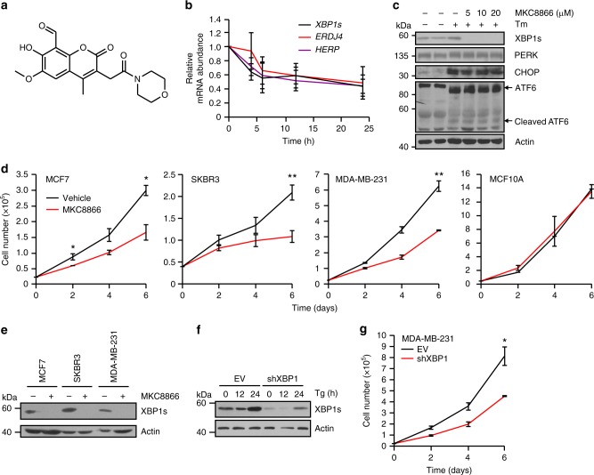
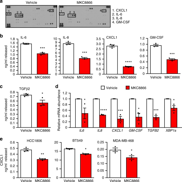
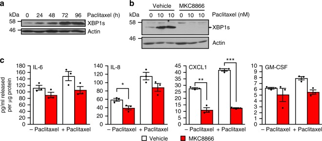
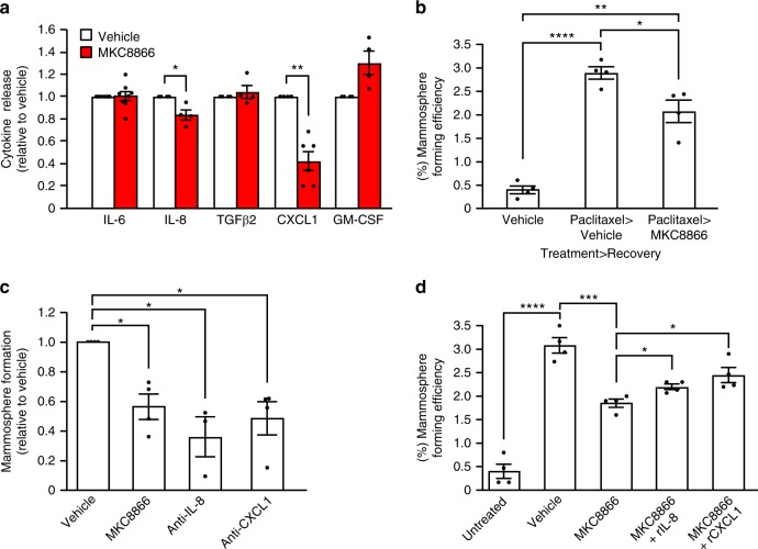
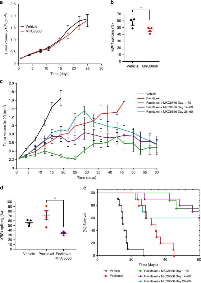
Triple-negative breast cancer (TNBC) lacks targeted therapies and has a worse prognosis than other breast cancer subtypes, underscoring an urgent need for new therapeutic targets and strategies. IRE1 is an endoplasmic reticulum (ER) stress sensor, whose activation is predominantly linked to the resolution of ER stress and, in the case of severe stress, to cell death. Here we demonstrate that constitutive IRE1 RNase activity contributes to basal production of pro-tumorigenic factors IL-6, IL-8, CXCL1, GM-CSF, and TGFβ2 in TNBC cells. We further show that the chemotherapeutic drug, paclitaxel, enhances IRE1 RNase activity and this contributes to paclitaxel-mediated expansion of tumor-initiating cells. In a xenograft mouse model of TNBC, inhibition of IRE1 RNase activity increases paclitaxel-mediated tumor suppression and delays tumor relapse post therapy. We therefore conclude that inclusion of IRE1 RNase inhibition in therapeutic strategies can enhance the effectiveness of current chemotherapeutics.


Conflict of interest statement

A.S., A.M.G., and E.C. are co-founders and shareholders of Cell Stress Discoveries Ltd. S.G., Q.Z., and J.B.P. are employees and shareholders of Fosun Orinove PharmaTech Inc. The remaining authors declare no competing interests.

Figures

Fig. 1
Fig. 1
Breast cancer cells exhibit constitutive IRE1 RNase activity. a Expression of spliced and unspliced XBP1 mRNA was assessed in a panel of breast cancer cell lines (MCF7, SKBR3, MDA-MB-231) and the non-tumorigenic breast epithelial cell line MCF10A via RT-PCR. Tunicamycin (Tm) (1 μg ml−1)-treated MCF10A cells act as a positive control for XBP1 splicing. GAPDH was used as a loading control. b Immunoblotting of XBP1s in a panel of breast cancer cell lines (MCF7, T47D, SKBR3, MDA-MB-231, and MDA-MB-468) and the non-tumorigenic breast epithelial cell line MCF10A. Tm (1 μg ml−1)-treated MCF10A cells act as a positive control for XBP1 splicing. Actin was used as a loading control. c Q-PCR quantification of the relative mRNA levels of spliced to total XBP1 in RNA samples obtained from basal-like tumor tissue (n = 5), luminal tumor tissue (n = 4), and tumor-associated normal (TAN) tissue (n = 4). Results shown for a and b are representative of three independent experiments. *P < 0.05 based on a pairwise single factor ANOVA tests comparing each tissue type. Error bars represent s.e.m.
Fig. 2
Fig. 2
Inhibition of IRE1 reduces breast cancer cell proliferation. a Chemical structure of MKC8866. b MDA-MB-231 cells were treated with 20 μM MKC8866 for 4, 6, 12, and 24 h after which RNA was extracted and levels of XBP1s, ERDJ4, and HERP mRNA transcripts quantified by Q-PCR (n = 4). c T47D cells were treated for 24 h with 1 μg ml−1 Tm alone or in combination with increasing concentrations of MKC8866 (5, 10, 20 μM) and cell lysates immunoblotted for XBP1s, PERK, CHOP, ATF6, and Actin. d MCF7 (n = 3), SKBR3 (n = 5), MDA-MB-231 (n = 2), and MCF10A (n = 3) cells were treated with 20 μM MKC8866 or an equal volume of DMSO and cell proliferation monitored by cell counts every second day for 6 days. e MCF7, SKBR3, and MDA-MB-231 cells were treated with 20 μM MKC8866 for 24 h and cell lysates immunoblotted for XBP1s and Actin. f Empty vector (EV) and XBP1shRNA MDA-MB-231 cells were treated for the indicated times with Tg (0.5 μM) after which expression of XBP1s and Actin was determined by immunoblotting. g Proliferation of empty vector (EV) and XBP1shRNA MDA-MB-231 cells was monitored by cell counts every second day for 6 days (n = 3). Results shown for c, e, and f are representative of three independent experiments. *P < 0.05, **P < 0.01 and ***P < 0.001, based on a Student’s t test. Error bars represent s.e.m.
Fig. 3
Fig. 3
IRE1 gene signature associates with basal-like breast cancers. a The putative IRE1 RNase-dependent gene signature was applied to a gene expression data set comprised of 27 breast cancer cell lines. Hierarchical clustering was performed and cell lines ranked based on their predicted IRE1 RNase activity. Expression across each gene (row) was centered and scaled so that mean expression is zero and standard deviation is one. Red indicates those genes with high expression and blue those with low expression relative to the mean. b IRE1 RNase-dependent gene signature was used to stratify 595 breast cancer gene expression data sets in TGCA. Cohorts with high and low IRE1 activity where identified and are represented as a heat map. Red indicates genes with high expression while blue those with low expression relative to the mean. c Pie charts depicting the breast cancer molecular sub-types (based on PAM50 classification) of IRE1 high and IRE1 low cohorts. NA indicates samples where PAM50 classification information was not available. d mRNA expression levels of IL6, IL8, CXCL1, GM-CSF, and TGFB2 in IRE1 high versus IRE1 low cohorts. Box plots show the median, 25th and 75th percentiles, and whiskers indicate the location of the minimum and maximum values for each of the IRE1 low (n = 79) and IRE1 high (n = 63) groups. e Correlation between immunohistochemistry staining intensity for XBP1s and IL-8 in TNBC human tumor sections (n = 16). f Correlation between immunohistochemistry staining intensity for XBP1s and CXCL1 in human TNBC tumor sections (n = 14). *P < 0.05, **P < 0.01, ***P < 0.001, and ****P < 0.0001, based on comparison of the two groups using a two-tailed t test with Welch’s correction. Error bars represent s.e.m.
Fig. 4
Fig. 4
IRE1 activity induces production of pro-inflammatory factors. ad MDA-MB-231 cells cultured in medium containing 2% serum were treated with 20 μM MKC8866 or vehicle alone for 48 h after which cells and conditioned medium were collected. a Conditioned medium was applied to a Human XL Cytokine Array. Expression profile of cytokines in vehicle alone versus MKC8866-conditioned medium was determined by chemiluminescence. b, c Cytokine secretion was quantified in conditioned medium using ELISAs selective for IL-6, IL-8, CXCL1, GM-CSF, and TGFβ2 (n = 3). d mRNA transcript levels of IL6, IL8, CXCL1, GM-CSF, TGFB2, and XBP1s were quantified by Q-PCR (n = 3). e CXCL1 quantification in conditioned medium collected from HCC1806, BT549, and MDA-MB-468 cells treated for 48 h in 2% serum-containing medium supplemented with vehicle alone or 20 μM MKC8866 (n = 3). Results shown for a are representative of two independent experiments. *P < 0.05, **P < 0.01, ***P < 0.001 and ****P < 0.0001, based on a Student’s t test. Error bars represent s.e.m.
Fig. 5
Fig. 5
Paclitaxel increases IRE1-dependent cytokine secretion. a MDA-MB-231 cells were treated with 10 nM paclitaxel for the indicated times, and cell lysates were immunoblotted for XBP1s and Actin. b MDA-MB-231 cells were treated with 10 nM paclitaxel in the presence of 20 μM MKC8866 or vehicle (DMSO) for 72 h, cell lysates were harvested and immunoblotted for XBP1s and Actin. c MDA-MB-231 cells were treated with 10 nM paclitaxel in combination with DMSO or 20 μM MKC8866 for 72 h in the presence of Boc-D-fmk (40 μM). Following treatment conditioned medium was collected and analyzed by ELISA for secretion of IL-6, IL-8, CXCL1, and GM-CSF. Cells were lysed and protein quantified (n = 3). Results shown for a and b are representative of three independent experiments. *P < 0.05, **P < 0.01, ***P < 0.001, based on a Student’s t test. Error bars represent s.e.m.
Fig. 6
Fig. 6
Paclitaxel alteration of secretome enhances mammosphere formation. ad MDA-MB-231 cells were treated with paclitaxel (10 nM) for 72 h, after which paclitaxel-containing medium was removed and cells were washed. a, b Cells were incubated for a further 72 h in fresh medium containing vehicle alone or MKC8866 (20 μM). a Conditioned medium was collected and levels of IL-6 (n = 7), IL-8 (n = 4), TGFβ2 (n = 4), CXCL1 (n = 6), and GM-CSF (n = 4) were quantified by ELISA. Cytokine release was normalized to that of vehicle-only treated controls. b After treatment, cells were seeded at equal densities onto low-adherence plates and mammospheres quantified after a further 5 days (n = 4). c, d Fresh medium containing vehicle alone, MKC8866 (20 μM), c neutralizing antibodies against CXCL1 (10 μg ml−1) or IL-8 (500 ng ml−1) (n = 4), d MKC8866 (20 μM) plus recombinant CXCL1 (rCXCL1, 500 pg ml−1) or recombinant IL-8 (rIL-8, 3 ng ml−1) was added and cells incubated for an additional 72 h. Cells were seeded at equal densities onto low-adherence plates and mammospheres quantified after a further 5 days (n = 4). *P < 0.05, **P < 0.01 and ***P < 0.001, based on a Student’s t test. Error bars represent s.e.m.
Fig. 7
Fig. 7
MKC8866 enhances the effectiveness of paclitaxel in vivo. Xenografts were established by subcutaneously injecting 5 × 106 MDA-MB-231 cells into the right flank of female athymic nude mice (Crl:NU(Ncr)-Foxn1nu, Charles River). When tumors were palpable (250 mm3) mice were randomized into groups and treatments initiated. a Vehicle-only versus MKC8866 (300 mg kg−1) daily via oral gavage. Tumor size was assessed every 2–3 days via caliper measurement and tumor volume calculated. By day 25, all tumors had reached their maximum permitted size (n = 10 mice per group). b Percentage XBP1 mRNA splicing was determined in vehicle-only versus MKC8866-treated xenografts (n = 4 per treatment group). c Paclitaxel was administered weekly at 10 mg kg−1 by intravenous injection, alone and in combination with MKC8866 administered daily at 300 mg kg−1 by oral gavage from day 1 to 60, from day 14 to 60, and from day 28 to 60. Tumor size was assessed every 2–3 days via caliper measurement and tumor volume calculated (n = 10 mice per group). d Percentage XBP1 mRNA splicing was determined in vehicle-only, MKC8866-treated and paclitaxel plus MKC8866-treated xenografts (n = 4 per treatment group). e Kaplan–Meier plot showing survival in animals administered with MKC8866 in combination with paclitaxel (for indicated times) compared to paclitaxel alone or vehicle alone. *P < 0.05, based on a Student’s t test. Error bars represent s.e.m.
Fig. 8
Fig. 8
MKC8866 reduces tumor regrowth post-paclitaxel withdrawal. Xenografts were established by subcutaneously injecting 5 × 106 MDA-MB-231 cells into the right flank of female athymic nude mice (Crl:NU(Ncr)-Foxn1nu, Charles River). When tumors were palpable (250 mm3) mice were randomized into groups and treatments initiated. Paclitaxel (7.5 mg kg−1 by intravenous injection) was administered every second day until day 10 (last dose indicated by the black arrow) as a single agent or in combination with MKC8866 (300 mg kg−1 by oral gavage). MKC8866 treatment was administered daily from day 1 to 28 (last dose indicated by the red arrow). Tumor size was assessed every 2–3 days via caliper measurement and tumor volume calculated (n = 10 mice per group). *P < 0.05, **P < 0.01 based on a Student’s t test. Error bars represent s.e.m.

References

    1. Szegezdi E, Logue SE, Gorman AM, Samali A. Mediators of endoplasmic reticulum stress-induced apoptosis. EMBO Rep. 2006;7:880–885. doi: 10.1038/sj.embor.7400779. - DOI - PMC - PubMed
    1. Korennykh A, Walter P. Structural basis of the unfolded protein response. Annu. Rev. Cell Dev. Biol. 2012;28:251–277. doi: 10.1146/annurev-cellbio-101011-155826. - DOI - PubMed
    1. Yoshida H, Matsui T, Yamamoto A, Okada T, Mori K. XBP1 mRNA is induced by ATF6 and spliced by IRE1 in response to ER stress to produce a highly active transcription factor. Cell. 2001;107:881–891. doi: 10.1016/S0092-8674(01)00611-0. - DOI - PubMed
    1. Lu Y, Liang FX, Wang X. A synthetic biology approach identifies the mammalian UPR RNA ligase RtcB. Mol. Cell. 2014;55:758–770. doi: 10.1016/j.molcel.2014.06.032. - DOI - PMC - PubMed
    1. Lee AH, Iwakoshi NN, Glimcher LH. XBP-1 regulates a subset of endoplasmic reticulum resident chaperone genes in the unfolded protein response. Mol. Cell. Biol. 2003;23:7448–7459. doi: 10.1128/MCB.23.21.7448-7459.2003. - DOI - PMC - PubMed

Publication types

MeSH terms