. 2018 Jul 10:2018:4924325.
doi: 10.1155/2018/4924325. eCollection 2018.

Improvement of Adipose Macrophage Polarization in High Fat Diet-Induced Obese GHSR Knockout Mice

Affiliations

Improvement of Adipose Macrophage Polarization in High Fat Diet-Induced Obese GHSR Knockout Mice

Fang Yuan et al. Biomed Res Int. .

Abstract

Purpose: Adipose tissue inflammation is the key linking obesity to insulin resistance. Over 50% of the interstitial cells in adipose tissue are macrophages, which produce inflammatory cytokines and therefore play an important role in the progression of insulin resistance. Within this classification view, macrophage biology is driven by two polarization phenotypes, M1 (proinflammatory) and M2 (anti-inflammatory). The unique functional receptor of ghrelin, growth hormone secretagogue receptor (GHSR), is a classic seven-transmembrane G protein-coupled receptor that is linked to multiple intracellular signaling pathways. Knockout of GHSR improves the obesity and glucose metabolic disorders, suggesting a crucial role of ghrelin activity in insulin resistance. Here, we discussed whether macrophage polarization phenotypes in adipose tissue were changed in GHSR knockout (GHSR-/-) mice.

Methods: GHSR-/- mice were fed with normal chow diet (NCD) or high fat diet (HFD). Markers of different macrophage polarization phenotypes were detected by real-time RT-PCR.

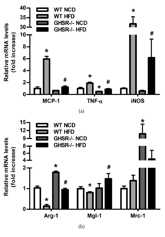
Results: The size of adipocytes decreased and interstitial cells, especially infiltrated macrophages, reduced in epididymal adipose tissue of GHSR-/- mice fed with HFD. Compared with wild type mice, the mRNA levels of inflammatory adipokines such as resistin, IL-6, and PAI-1 were significantly lower in epididymal adipose tissue of GHSR-/- mice, whereas anti-inflammatory adipokine, adiponectin, was significantly higher. M1 markers, MCP-1, TNF-α, and iNOS, were significantly lower in epididymal adipose tissue of GHSR-/- mice, whereas M2 markers, Arg-1, Mgl-1, were Mrc1, were significantly higher.

Conclusion: The GHSR-/- mice fed with HFD showed suppressed adipose inflammation, reduced macrophage infiltration, and enhanced M2 polarization of macrophages in adipose tissue, which improved insulin sensitivity.


Figures

Figure 1
Figure 1
Improvement of glucose metabolism in GHSR knockout mice. (a) Body weight of GHSR knockout (GHSR-/-) mice and wild type (WT) mice fed with different diet. (b) The average daily food intake of GHSR-/- mice and wild type (WT) mice fed with normal chow diet or high fat diet for 12 weeks. (c) The average daily water intake of GHSR-/- mice and wild type mice fed with normal chow diet or high fat diet. (d) Results of glucose tolerance test using GHSR knockout mice and wild type mice fed with normal chow diet (NCD) or high fat diet (HFD) for 12 weeks. All results were expressed as means±SEM. (e) Western blotting results from GHSR knockout mice and wild type mice fed with normal chow diet or high fat diet for 12 weeks. Insulin at a dose of 2 IU/kg was injected intraperitoneally 15 min before harvest of the adipose tissue; phospho-Akt (Ser473) and Akt in adipose tissue were detected using specific antibodies. Total Akt level was used as internal control. Quantification of image analysis of p-Akt/Akt was expressed as means±SEM. denotes P<0.05 compared with wild type mice fed with NCD; # denotes P<0.05 compared with WT mice fed with HFD.
Figure 2
Figure 2
Improvement of adipose tissue inflammation in GHSR knockout mice. (a) Expression of inflammatory adipokines in epididymal adipose tissue of GHSR knockout mice, such as anti-inflammatory adipokine adiponectin and proinflammatory adipokines resistin, IL-6, and PAI-1. Relative mRNA levels were normalized to the levels for wild type mice fed with normal chow diet. Data are means±SEM, n=5. (b) Adipocytes volume and interstitial cell infiltration detected by H&E staining. (c) Macrophage infiltration detected by immunofluorescence histochemistry staining of Mac-3. denotes P<0.05 compared with WT mice fed with NCD; # denotes P<0.05 compared with WT mice fed with HFD.
Figure 3
Figure 3
Macrophage polarization in epididymal adipose tissue of GHSR knockout mice. The mRNAs were extracted from the epididymal adipose tissue harvested from the GHSR knockout mice and wild type mice fed with different diet. Real-time PCR was performed to evaluate the expression of macrophage specific markers. (a) M1 markers MCP-1, TNF-α, iNOS and (b) M2 markers Arg-1, Mgl-1, Mrc1 were detected. Data are means±SEM n=5. denotes P<0.05 compared with WT mice fed with NCD; # denotes P<0.05 compared with WT mice fed with HFD.
Figure 4
Figure 4
Acyl ghrelin influences the effect of LPS or IL-4 on RAW264.7 cell polarization. The mRNAs were extracted from the RAW 264.7 pretreated with acyl ghrelin for 30 minutes then for 6 hours with LPS (10 ng/ml) (a) or IL-4 (10 ng/ml) (b). Real-time PCR was performed to evaluate the expression of macrophage specific markers Arg-1 and MCP-1. Data are means±SEM from 3 separate experiments. denotes P<0.05 compared with control group; # denotes P<0.05 compared with cells treated with LPS or IL-4 alone.

References

    1. Kojima M., Hosoda H., Date Y., Nakazato M., Matsuo H., Kangawa K. Ghrelin is a growth-hormone-releasing acylated peptide from stomach. Nature. 1999;402(6762):656–660. doi: 10.1038/45230. - DOI - PubMed
    1. Koyama K.-I., Yasuhara D., Nakahara T., et al. Changes in acyl ghrelin, des-acyl ghrelin, and ratio of acyl ghrelin to total ghrelin with short-term refeeding in female inpatients with restricting-type anorexia nervosa. Hormone and Metabolic Research. 2010;42(8):595–598. doi: 10.1055/s-0030-1252017. - DOI - PubMed
    1. Gutierrez J. A., Solenberg P. J., Perkins D. R., et al. Ghrelin octanoylation mediated by an orphan lipid transferase. Proceedings of the National Acadamy of Sciences of the United States of America. 2008;105(17):6320–6325. doi: 10.1073/pnas.0800708105. - DOI - PMC - PubMed
    1. Yang J., Brown M. S., Liang G., Grishin N. V., Goldstein J. L. Identification of the Acyltransferase that Octanoylates Ghrelin, an Appetite-Stimulating Peptide Hormone. Cell. 2008;132(3):387–396. doi: 10.1016/j.cell.2008.01.017. - DOI - PubMed
    1. Delhanty P. J. D., Sun Y., Visser J. A., et al. Unacylated ghrelin rapidly modulates lipogenic and insulin signaling pathway gene expression in metabolically active tissues of GHSR deleted mice. PLoS ONE. 2010;5(7) doi: 10.1371/journal.pone.0011749.e11749 - DOI - PMC - PubMed

Substances

LinkOut - more resources