Review
. 2018 Oct;9(5):709-719.
doi: 10.1007/s13244-018-0647-9. Epub 2018 Aug 15.

Radiological findings of complications after lung transplantation

Affiliations
Review

Radiological findings of complications after lung transplantation

Céline Habre et al. Insights Imaging. 2018 Oct.

Abstract

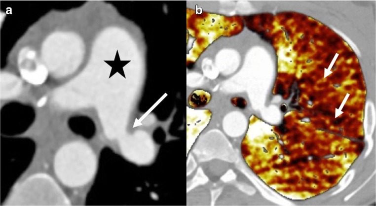
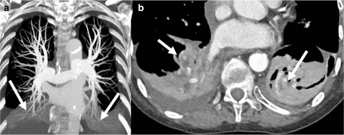
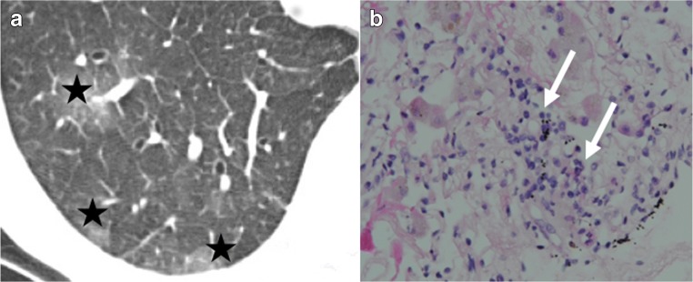
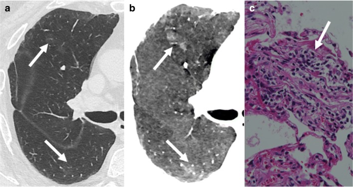
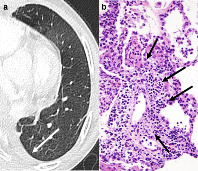
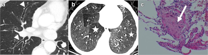
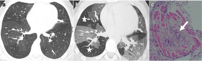
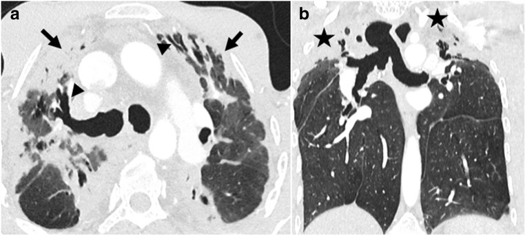
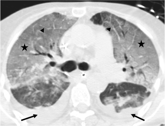
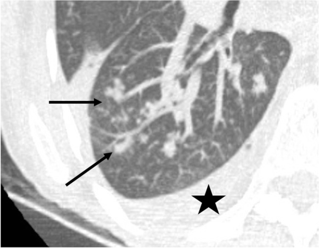
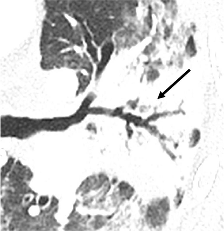
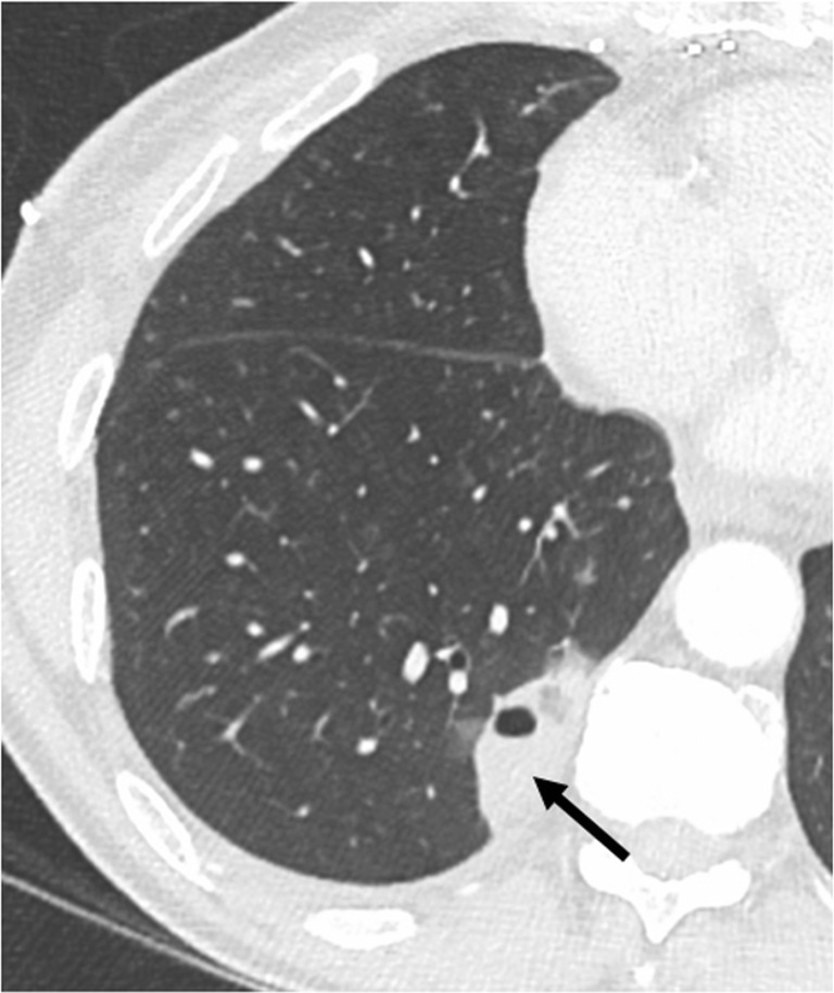
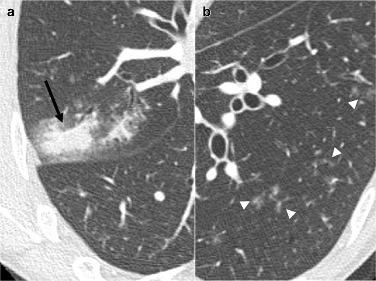
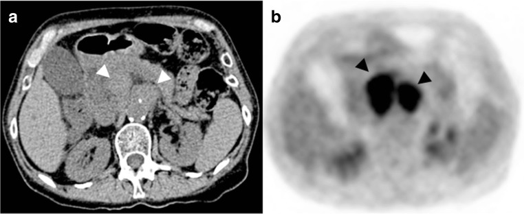
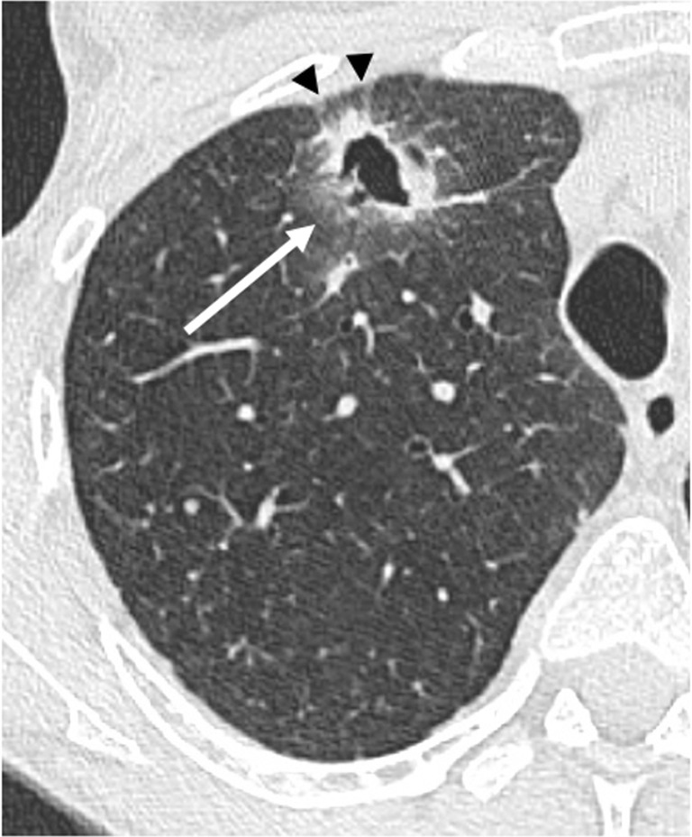
Complications following lung transplantation may impede allograft function and threaten patient survival. The five main complications after lung transplantation are primary graft dysfunction, post-surgical complications, alloimmune responses, infections, and malignancy. Primary graft dysfunction, a transient ischemic/reperfusion injury, appears as a pulmonary edema in almost every patient during the first three days post-surgery. Post-surgical dysfunction could be depicted on computed tomography (CT), such as bronchial anastomosis dehiscence, bronchial stenosis and bronchomalacia, pulmonary artery stenosis, and size mismatch. Alloimmune responses represent acute rejection or chronic lung allograft dysfunction (CLAD). CLAD has three different forms (bronchiolitis obliterans syndrome, restrictive allograft syndrome, acute fibrinoid organizing pneumonia) that could be differentiated on CT. Infections are different depending on their time of occurrence. The first post-operative month is mostly associated with bacterial and fungal pathogens. From the second to sixth months, viral pneumonias and fungal and parasitic opportunistic infections are more frequent. Different patterns according to the type of infection exist on CT. Malignancy should be depicted and corresponded principally to post-transplantation lymphoproliferative disease (PTLD). In this review, we describe specific CT signs of these five main lung transplantation complications and their time of occurrence to improve diagnosis, follow-up, medical management, and to correlate these findings with pathology results. KEY POINTS: • The five main complications are primary graft dysfunction, surgical, alloimmune, infectious, and malignancy complications. • CT identifies anomalies in the setting of unspecific symptoms of lung transplantation complications. • Knowledge of the specific CT signs can allow a prompt diagnosis. • CT signs maximize the yield of bronchoscopy, transbronchial biopsy, and bronchoalveolar lavage. • Radiopathological correlation helps to understand CT signs after lung transplantation complications.

Keywords: Lung transplant complications; Radiological findings.


Conflict of interest statement

The local institutional review board approved the study.

Figures

Fig. 1
Fig. 1
Onset of complications following lung transplantation. Adapted from Ng et al. 2009 [4]. CLAD: chronic lung allograft dysfunction; PGD: primary graft dysfunction; PTLD: post-transplantation lymphoproliferative disease; D: day; W: week; M: month; Y: year
Fig. 2
Fig. 2
Primary graft dysfunction. Septal, scissural, and peribronchovascular thickening (arrowheads) and pleural effusions (stars)
Fig. 3
Fig. 3
Bronchial anastomosis dehiscence. Air collection anterior to the right bronchial suture (arrow) (a). Indirect signs include persistent pneumothorax (arrow) and new subcutaneous emphysema (arrowheads) (b)
Fig. 4
Fig. 4
Bronchial stenosis. Dyspnea and recurrent infections at 6 months after lung transplantation. Narrowing of the anastomosis of the right main bronchus with minimal intensity projection (arrow)
Fig. 5
Fig. 5
Pulmonary artery stenosis. Dyspnea and chest pain 3 months after surgery. Stenosis of the left PA (arrow) responsible for dilatation of the main PA (star) (a) and, as consequences, a wide hypoperfusion of the left lung in the pale yellow color area on the perfusion map (arrows) (b)
Fig. 6
Fig. 6
Donor–recipient size mismatch. Persisting dyspnea. Atelectasis of lower lobes (arrows in a) with comet-tail sign (arrows in b)
Fig. 7
Fig. 7
Acute rejection A1. Ground-glass opacities (stars) and interlobular thickening (arrowheads) (a) in minimal acute rejection, characterized by sparse perivascular mononuclear infiltrates (arrows) (b)
Fig. 8
Fig. 8
Acute rejection A2. Subtle ground-glass opacities (arrows in a) better detected with minimal intensity projection (arrows in b) in mild acute rejection, with more frequent perivascular mononuclear infiltrates and endothelialitis (arrow in c)
Fig. 9
Fig. 9
Acute rejection A3. New pleural effusion (arrow) (a) as the only finding in moderate acute rejection, histologically defined as dense perivascular mononuclear infiltrates with peribronchiolar and airspaces extension (arrows) (b)
Fig. 10
Fig. 10
Bronchiolitis obliterans syndrome (BOS). Chronic dyspnea with obstructive pattern at functional tests. Bronchiectases, bronchial thickening (arrowheads) (a) and mosaic perfusion on minimal intensity projection reconstruction (radiolucent areas; stars) (b) in constrictive bronchiolitis, characterized by submucosa fibrosis with mononuclear infiltrates of the small airways (arrow) (c)
Fig. 11
Fig. 11
BOS. Chronic dyspnea and cough. Normal parenchyma on inspiration acquisition (a) followed by air-trapping on expiration acquisition (stars) (b) in constrictive bronchiolitis, characterized by submucosa fibrosis (arrow) (c)
Fig. 12
Fig. 12
Restrictive allograft syndrome (RAS). Dyspnea and irreversible decline in forced expiratory volume (FEV) and total lung capacity (TLC). Peripheral condensations and subpleural thickening (arrows), bronchiectasis (arrowheads) (a) and volume loss with upper lobe predominance (stars) (b). RAS was confirmed by biopsy
Fig. 13
Fig. 13
Acute fibrinoid allograft pneumonia (AFOP). Acute shortness of breath. Intralobular septal thickening and extensive ground-glass opacities (stars), interlobular thickening (arrowheads), and pleural effusions (arrows), confirmed by histology
Fig. 14
Fig. 14
Pseudomonas aeruginosa. Productive cough and high-grade fever. Dense tree-in-bud centrilobular nodules on maximal intensity projection reconstruction (arrows) and pleural effusion (star). Pseudomonas aeruginosa was confirmed by sputum culture
Fig. 15
Fig. 15
Staphylococcus epidermidis. Sepsis. Consolidation with air bronchogram (arrow)
Fig. 16
Fig. 16
Lung abscess. Fever of unknown origin and asthenia. Consolidation with air–fluid level (arrow) suggestive of lung abscess. Methicillin-susceptible S. aureus (MSSA) infection was confirmed by bronchoalveolar lavage
Fig. 17
Fig. 17
Angioinvasive Aspergillus fumigatus. General alteration and productive cough. Solid nodule with peripheral ground-glass opacities termed as the “halo sign” (arrow) (a). Pathology showed branching filaments (arrows), consistent with angioinvasive aspergillosis (b)
Fig. 18
Fig. 18
Parainfluenzae pneumonia. Sepsis. Segmental consolidation of the right upper lobe (arrow) (a) and left basal ground-glass centrilobular micronodulation (arrowheads) (b)
Fig. 19
Fig. 19
Post-transplantation lymphoproliferative disease (PTLD). Weight loss and general alteration. Voluminous retroperitoneal lymphadenopathies on non-enhanced abdominal CT (arrowheads) (a) with increased metabolism on FDG-PET CT scanning (arrowheads) (b)
Fig. 20
Fig. 20
Primary epidermoid lung carcinoma. Asymptomatic, cavitated nodule on follow-up chest X-ray. Cavitated nodule with spiculated margins (arrow) and pleural traction (arrowheads)

References

    1. Yusen RD, Edwards LB, Dipchand AI, et al. The registry of the International Society for Heart and Lung Transplantation: thirty-third adult lung and heart-lung transplant report-2016; focus theme: primary diagnostic indications for transplant. J Heart Lung Transplant. 2016;35:1170–1184. doi: 10.1016/j.healun.2016.09.001. - DOI - PubMed
    1. Lund LH, Khush KK, Cherikh WS, et al. The registry of the International Society for Heart and Lung Transplantation: thirty-fourth adult heart transplantation report-2017; focus theme: allograft ischemic time. J Heart Lung Transplant. 2017;36:1037–1046. doi: 10.1016/j.healun.2017.07.019. - DOI - PubMed
    1. Kroegel C, Hekmat K, Möser A, Happe J, Walther R, Reissig A. Airway complications following lung transplantation—clinic, diagnosis, and interventional management. Pneumologie. 2011;65:293–307. doi: 10.1055/s-0030-1255764. - DOI - PubMed
    1. Ng YL, Paul N, Patsios D, et al. Imaging of lung transplantation: review. AJR Am J Roentgenol. 2009;192:S1–S13. doi: 10.2214/AJR.08.1544. - DOI - PubMed
    1. Fisher CE, Preiksaitis CM, Lease ED, et al. Symptomatic respiratory virus infection and chronic lung allograft dysfunction. Clin Infect Dis. 2016;62:313–319. doi: 10.1093/cid/civ871. - DOI - PMC - PubMed

LinkOut - more resources