Review
. 2019;20(2):131-145.
doi: 10.1631/jzus.B1800084. Epub 2018 Aug 4.

Bone morphogenetic proteins and inner ear development

Affiliations
Review

Bone morphogenetic proteins and inner ear development

Jiao-Yao Ma et al. J Zhejiang Univ Sci B. 2019.

Abstract

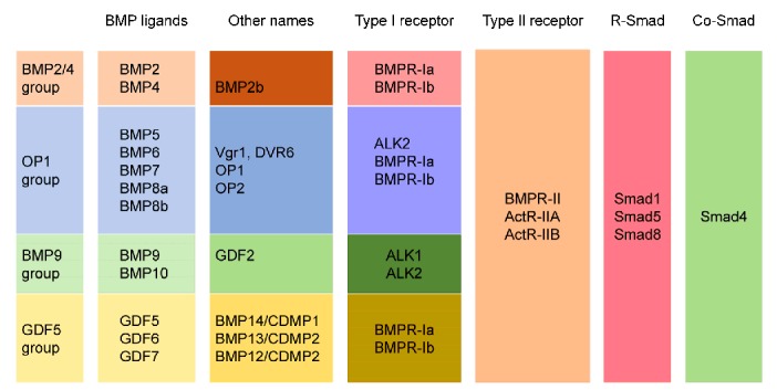
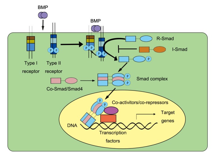
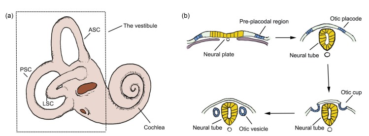
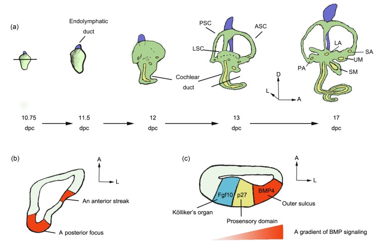
Bone morphogenetic proteins (BMPs) are the largest subfamily of the transforming growth factor-β superfamily, and they play important roles in the development of numerous organs, including the inner ear. The inner ear is a relatively small organ but has a highly complex structure and is involved in both hearing and balance. Here, we discuss BMPs and BMP signaling pathways and then focus on the role of BMP signal pathway regulation in the development of the inner ear and the implications this has for the treatment of human hearing loss and balance dysfunction.

Keywords: Bone morphogenetic protein (BMP) signaling; Development; Inner ear; Hearing; Balance.


Conflict of interest statement

Compliance with ethics guidelines: Jiao-yao MA, Dan YOU, Wen-yan LI, Xiao-ling LU, Shan SUN, and Hua-wei LI declare that they have no conflict of interest.

This review does not contain any studies with human or animal subjects performed by any of the authors.

Figures

Fig. 1
Fig. 1
BMPs, BMP receptors, and activated Smads Based on their function and structure, BMPs are divided into four subgroups: (1) BMPs 2 and 4; (2) BMPs 5, 6, 7, 8a, and 8b; (3) BMPs 9 and 10; and (4) BMPs 12, 13, and 14. In addition, the related BMP receptors, Smad proteins, and some other names for the BMP ligands are listed. BMP, bone morphogenetic protein; OP, osteogenic protein; GDF, growth differentiation factor; Vgr, vegetal related; DVR, decapentaplegic vegetal related; CDMP, cartilage-derived morphogenetic protein; BMPR, BMP receptor; ALK, activin receptor-like kinase; ActR-II, activin type II receptor
Fig. 2
Fig. 2
Smad-dependent BMP signaling BMP ligands interact with the BMP receptor complex consisting of type І and type II receptors (BMPR-І and BMPR-ІI, respectively). After the interaction, BMPR-І phosphorylates BMPR-II and leads to the phosphorylation of R-Smads (Smad1/Smad5/Smad8). This activation allows the formation of Smad complexes consisting of R-Smads (Smad1/Smad5/Smad8) and Co-Smad (Smad4). The Smad complexes then translocate into the nucleus where they induce transcription of the target genes. R-Smad, receptor-regulated Smad; Co-Smad, common-mediator Smad; I-Smad, inhibitory Smad
Fig. 3
Fig. 3
Inner ear and the induction of the otocyst (a) The inner ear consists of the cochlea, which is involved in hearing, and the vestibule, which is involved in balance. (b) The pre-placodal region is a zone of ectoderm that lies lateral to the neural plate. It gives rise to all sensory placodes in the head, including the otic placode. The placode invaginates into the otic cup, which later closes to become the otic vesicle/otocyst. ASC, anterior semicircular canal; LSC, lateral semicircular canal; PSC, posterior semicircular canal
Fig. 4
Fig. 4
Specification of the inner ear (a) The development of the mouse inner ear from 10.75 to 17 d past coitum (dpc). (b) In the mouse otocyst, BMP4 expression was detected in two regions at 10.75 dpc—an anterior streak and a posterior focus. (c) In the mouse cochlea, a gradient of BMP signaling was detected along the abneural–neural axis at 13.5 dpc. The BMP4 expression is high in the outer sulcus and low in Kölliker’s organ. ASC, anterior semicircular canal; LSC, lateral semicircular canal; PSC, posterior semicircular canal; LA, lateral ampulla; PA, posterior ampulla; SA, superior ampulla; SM, saccular macula; UM, utricular macula; A, anterior; D, dorsal; L, lateral

References

    1. Abelló G, Khatri S, Radosevic M, et al. Independent regulation of Sox3 and Lmx1b by FGF and BMP signaling influences the neurogenic and non-neurogenic domains in the chick otic placode. Dev Biol. 2010;339(1):166–178. doi: 10.1016/j.ydbio.2009.12.027. - DOI - PubMed
    1. Alsina B, Giraldez F, Pujades C. Patterning and cell fate in ear development. Int J Dev Biol. 2009;53(8-10):1503–1513. doi: 10.1387/ijdb.072422ba. - DOI - PubMed
    1. Alvarez IS, Navascués J. Shaping, invagination, and closure of the chick embryo otic vesicle: scanning electron microscopic and quantitative study. Anat Rec. 1990;228(3):315–326. doi: 10.1002/ar.1092280311. - DOI - PubMed
    1. Avsian-Kretchmer O, Hsueh AJ. Comparative genomic analysis of the eight-membered ring cystine knot-containing bone morphogenetic protein antagonists. Mol Endocrinol. 2004;18(1):1–12. doi: 10.1210/me.2003-0227. - DOI - PubMed
    1. Bai S, Cao X. A nuclear antagonistic mechanism of inhibitory Smads in transforming growth factor-β signaling. J Biol Chem. 2002;277(6):4176–4182. doi: 10.1074/jbc.M105105200. - DOI - PubMed

MeSH terms

LinkOut - more resources