. 2018 Aug;97(33):e11848.
doi: 10.1097/MD.0000000000011848.

Rhabdomyolysis revisited: Detailed analysis of magnetic resonance imaging findings and their correlation with peripheral neuropathy

Affiliations

Rhabdomyolysis revisited: Detailed analysis of magnetic resonance imaging findings and their correlation with peripheral neuropathy

Jun Ho Kim et al. Medicine (Baltimore). 2018 Aug.

Abstract

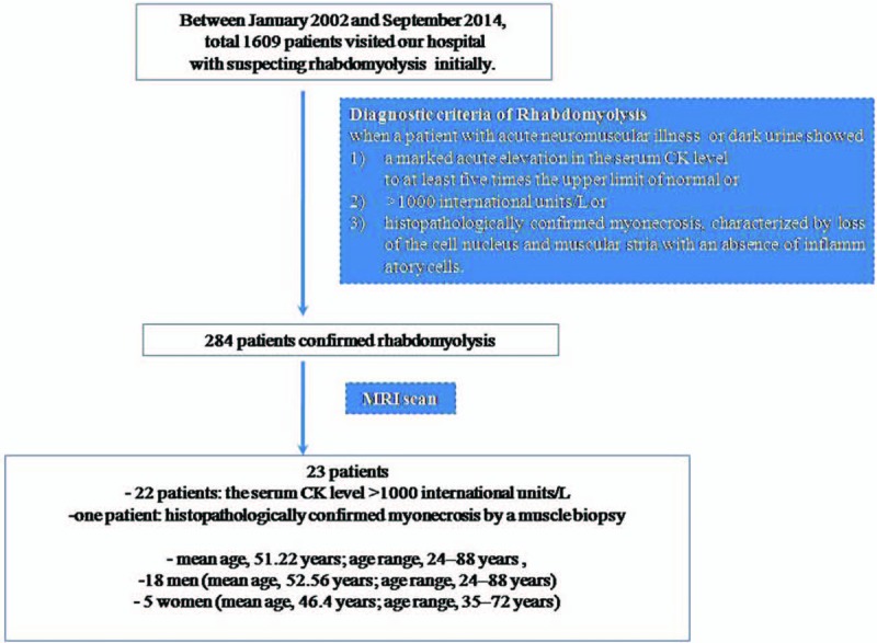
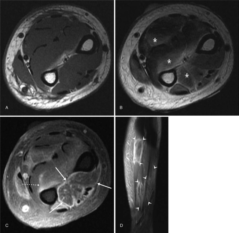
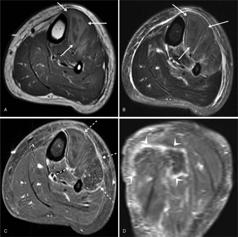
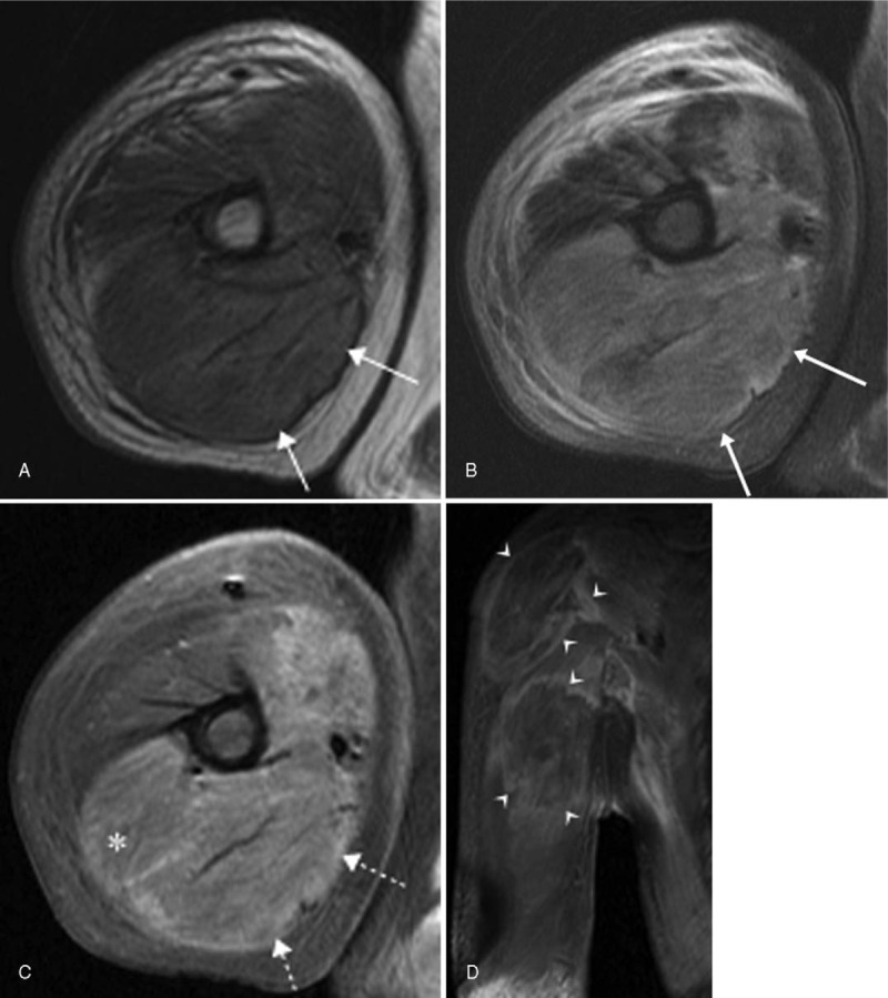
The objective is to evaluate the magnetic resonance imaging (MRI) findings in rhabdomyolysis in detail and determine their correlation with the development of peripheral neuropathy.Magnetic resonance images for 23 patients with confirmed rhabdomyolysis with (n = 11) or without (n = 12) peripheral neuropathy were retrospectively reviewed for the signal intensity on T1- and T2-weighted images, intramuscular hemorrhage, enhancement pattern, shape and margin in the longitudinal plane, edema in the deep fascia and overlying subcutaneous layer, multiplicity, and bilateral limb involvement. The collected data were statistically analyzed and the relationship between the imaging findings and the development of peripheral neuropathy was determined.Abnormal signal intensities on T1- or T2-weighted images were observed for all patients except one. Fourteen patients (60.9%) showed intramuscular hemorrhage. Stippled enhancement (11/23; 47.8%) was the most common enhancement pattern. Nineteen patients (86.4%) showed a well-defined rectangular shape with a ragged margin in the longitudinal plane. The affected muscle volume usually increased (17/23; 73.9%), with edema in the deep fascia and the overlying subcutaneous layer (13/23; 56.5%). Multiplicity within a muscle, compartment, and limb was observed in 7 (31.8%), 18 (81.8%), and 16 (72.7%) patients, respectively. Bilateral involvement was observed in 7 patients (30.4%). Only multiplicity within a compartment showed a statistically significant correlation with peripheral neuropathy development.Common MRI findings in rhabdomyolysis include intramuscular hemorrhage, stippled enhancement, a well-defined rectangular shape with a ragged margin in the longitudinal plane, and multiplicity. Multiplicity within a compartment may be a predictor of the development of peripheral neuropathy.


Conflict of interest statement

The authors have no conflicts of interest to disclose.

Figures

Figure 1
Figure 1
Flowchart showing the study selection procedure.
Figure 2
Figure 2
Drawings illustrating the enhancement patterns for major muscles affected by rhabdomyolysis. A, The enhancement pattern appears to be the same for the affected and unaffected muscle (iso-enhancement). B, Diffuse increased enhancement of the affected muscle relative to the adjacent normal muscle. C, Peripheral enhancement with central dot-like or linear streaky enhancement (so-called stippled enhancement). D, Central nonenhancing portion with peripheral enhancement.
Figure 3
Figure 3
Drawings illustrating the shape and margin of rhabdomyolysis lesions in the longitudinal plane (coronal or sagittal images) of contrast-enhanced fat-suppressed T1-weighted sequences. A, Ill-defined patchy shape. B, Well-defined round shape. C, Well-defined rectangular shape with ragged margin.
Figure 4
Figure 4
Axial T1-weighted (A), T2-weighted (B), contrast-enhanced, fat-suppressed T1-weighted (C), sagittal contrast-enhanced, fat-suppressed T1-weighted (D) images for a 41-year-old man with rhabdomyolysis due to drug intoxication. On a T1-weighted image (A), all the muscles show iso-signal intensity relative to the adjacent normal muscle (A). On a T2-weighted image (B), the affected areas of the muscles show homogeneous high signal intensity (asterisks). A contrast-enhanced, fat-suppressed T1-weighted image (C) shows peripheral enhancement with a central dot-like or linear streaky enhancement (so-called stippled enhancement) (arrows) of the muscles in the dorsal compartment, whereas some muscles in the deep volar compartment show diffuse increased enhancement (dashed arrows). On a sagittal contrast-enhanced, fat-suppressed T1-weighted image (D), the lesions show a well-defined rectangular shape with a ragged margin (arrowheads).
Figure 5
Figure 5
Axial T1-weighted (A), fat-suppressed T2-weighted (B), axial contrast-enhanced fat-suppressed T1-weighted (C), coronal contrast-enhanced fat-suppressed T1 weighted (D) images for a 46-year-old woman with rhabdomyolysis in the left lower leg due to drug intoxication. Heterogeneous signal intensity in the anterior compartment of the lower leg, with increased muscle volume, can be seen on T1-weighted (A) and fat-suppressed T2-weighted (B) images. The area showing high signal intensity on the T1-weighted and fat-suppressed T2-weighted (arrows) images is interpreted to be intramuscular hemorrhage. On an axial contrast-enhanced, fat-suppressed T1-weighted image (C), peripheral enhancement with a central non-enhancing portion can be seen (dashed arrows). On a coronal contrast-enhanced, fat-suppressed T1-weighted image (D), the lesion shows a well-defined rectangular shape with a ragged margin (arrowheads).
Figure 6
Figure 6
Axial T1-weighted (A), fat-suppressed T2-weighted (B), contrast-enhanced, fat-suppressed T1-weighted (C), coronal contrast-enhanced, fat-suppressed T1-weighted (D) images fora 43-year-old man with rhabdomyolysis caused by excessive exercise. Heterogeneous signal intensity in the posterior compartment and medial portion of the anterior compartment in the upper arm, with increased muscle volume (arrows), can be seen on T1-weighted (A) and fat-suppressed T2-weighted (B) images. Note the edema in the subcutaneous layer. On a contrast-enhanced, fat-suppressed T1-weighted image (C), diffuse enhancement is noted in not only the area with heterogeneous high signal intensity (dashed arrows) but also the area with iso-signal intensity on aprecontrastT1-weighted image (∗). On a coronal contrast-enhanced, fat-suppressed T1-weighted image (D), the lesions show a well-defined rectangular shape with a ragged margin (arrowheads).

Similar articles

Cited by

References

    1. Thomas MA, Ibels LS. Rhabdomyolysis and acute renal failure. Aust N Z J Med 1985;15:623–8. - PubMed
    1. McDonald S, Bearcroft P. Compartment syndromes. Semin MusculoskeletRadiol 2010;14:236–44. - PubMed
    1. Kim JY, Lee JW, Cha SO, et al. Compressive radial neuropathy developed under a fibrotic band associated with rhabdomyolysis and successfully treated with surgery. Ann Rehabil Med 2014;38:421–6. - PMC - PubMed
    1. Moratalla MB, Braun P, Fornas GM. Importance of MRI in the diagnosis and treatment of rhabdomyolysis. Eur J Radiol 2008;65:311–5. - PubMed
    1. Delaney-Sathy LO, Fessell DP, Jacobson JA, et al. Sonography of diabetic muscle infarction with MR imaging, CT, and pathologic correlation. AJR Am J Roentgenol 2000;174:165–9. - PubMed

Publication types

MeSH terms