

Brown tumor of multiple facial bones associated with primary hyperparathyroidism: A clinical case report
- PMID: 30113484
- PMCID: PMC6112971
- DOI: 10.1097/MD.0000000000011877
Brown tumor of multiple facial bones associated with primary hyperparathyroidism: A clinical case report
Abstract
Rationale: Only 4.5% of brown tumors involve facial bones; of these, solitary bone involvement is usual. Brown tumors of multiple facial bones are extremely rare. Here, we report the case of a brown tumor of multiple facial bones initially misdiagnosed as an odontogenic cyst.
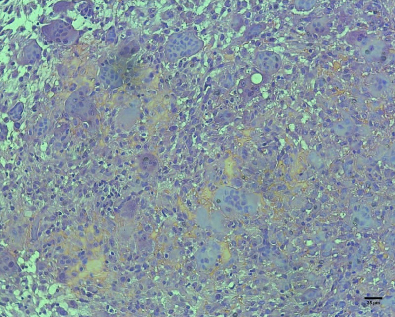
Patient concerns: A pregnant 26-year-old woman was referred to our hospital with painful swelling of multiple facial bones, anemia, urinary calculi, marasmus, and a history of multiple bone fractures. Laboratory examination revealed an elevated serum calcium level of 3.09 mmol/L (normal range: 2.0-2.8 mmol/L) and a low phosphorus level of 0.62 mmol/L (normal range: 0.81-1.65 mmol/L). The serum alkaline phosphatase concentration was 397 IU/L (normal range: 24-82 IU/L) and parathyroid hormone level was 267 pg/mL (normal range: 14-72 pg/mL). Cone beam computed tomography revealed multiple ossifying fibromas of the maxilla and mandible. Incisional biopsy revealed abundant spindle cells with areas of hemorrhage and haphazardly arranged diffuse multinucleated giant cells.
Diagnoses: The patient was diagnosed with primary hyperparathyroidism (HPT).
Interventions: She was treated by parathyroidectomy.
Outcomes: The multiple osteitis fibrosa cystica gradually resolved as bone re-mineralized. The patient has been followed up for 2 years without evidence of tumor recurrence.
Lessons: As multiple osteolytic lesions of facial bones can be caused by primary HPT, serum calcium and parathyroid hormone assays should be performed routinely when investigating these lesions.
Conflict of interest statement
The authors declare no conflicts of interest.
Figures


References
-
- Bilezikian JP, Silverberg SJ. Clinical practice. Asymptomatic primary hyperparathyroidism. N Engl J Med 2004;350:1746–51. - PubMed
-
- Silverberg SJ, Bilezikian JP. Incipient” primary hyperparathyroidism: a “forme fruste” of an old disease. J Clin Endocrinol Metab 2003;88:5348–52. - PubMed
-
- Syed H, Khan A. Primary hyperparathyroidism: diagnosis and management in 2017. Pol Arch Intern Med 2017;127:438–41. - PubMed
Publication types
MeSH terms
LinkOut - more resources
Full Text Sources
Other Literature Sources
Medical
Research Materials
