. 2019 Feb;67(2):107-116.
doi: 10.1369/0022155418793588. Epub 2018 Aug 16.

Expression of Non-collagenous Bone Matrix Proteins in Osteoblasts Stimulated by Mechanical Stretching in the Cranial Suture of Neonatal Mice

Affiliations

Expression of Non-collagenous Bone Matrix Proteins in Osteoblasts Stimulated by Mechanical Stretching in the Cranial Suture of Neonatal Mice

Mika Ikegame et al. J Histochem Cytochem. 2019 Feb.

Abstract

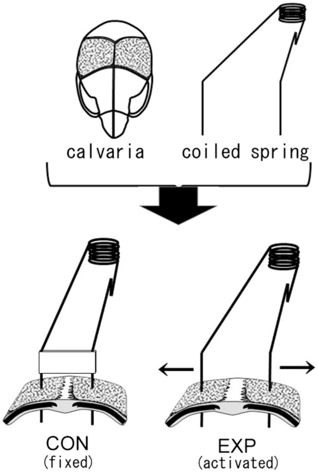
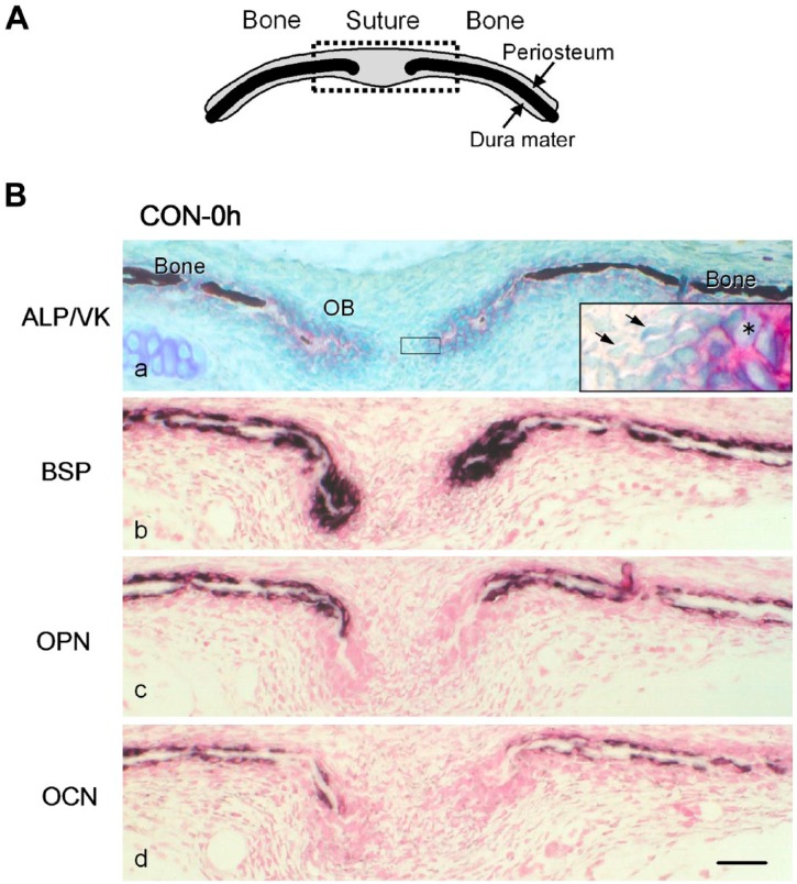
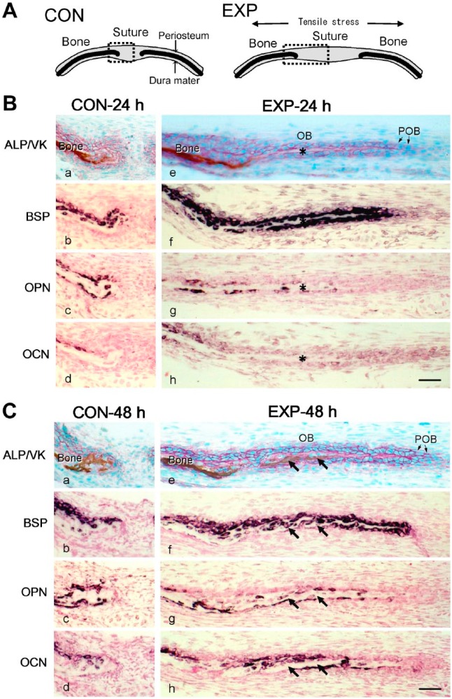
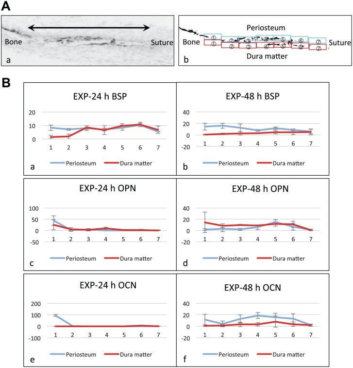
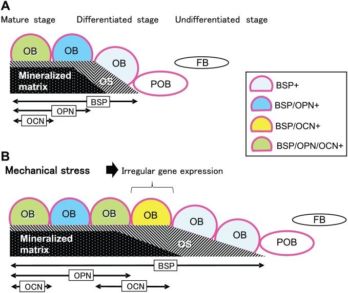
We investigated the influence of mechanical stretching on the genetic expression pattern of non-collagenous bone matrix proteins in osteoblasts. The cranial sutures of neonatal mice were subjected to ex vivo mechanical stretching. In the non-stretched control group, as osteoblast differentiation progressed, the successive genetic expression of bone sialoprotein (BSP), osteopontin (OPN), and osteocalcin (OCN) was detected using in situ hybridization, in that order. In the stretched group, the sutures were widened, and after 24 hr of cultivation, a large number of osteoblasts and abundant new osteoid were observed on the borders of the parietal bones. All new osteoblasts expressed BSP and some of them expressed OPN, but very few of them expressed OCN. After 48 hr, more extensive presence of osteoid was noted on the borders of the parietal bones, and this osteoid was partially mineralized; all osteoblasts on the osteoid surface expressed BSP, and more osteoblasts expressed OPN than those after 24 hr cultivation. Surprisingly, many of the osteoblasts that did not express OPN, expressed OCN. This suggests that when osteoblast differentiation is stimulated by mechanical stress, the genetic expression pattern of non-collagenous proteins in the newly differentiated osteoblasts is affected.

Keywords: mechanical loading; non-collagenous bone proteins; ossification; sagittal sutures.


Conflict of interest statement

Competing Interests: The author(s) declared no potential conflicts of interest with respect to the research, authorship, and/or publication of this article.

Figures

Figure 1.
Figure 1.
Schematic representation of a mouse calvaria and coiled springs. In the control group (CON), a fixed coiled spring maintains static width in the suture between parietal bones (dotted part of the calvaria). In the experimental group (EXP), an activated coiled spring exerts tensile stress in the sagittal suture and enlarges the suture in the direction of the arrows.
Figure 2.
Figure 2.
(A) A schematic frontal view of the sagittal suture and parietal bones. The upper and lower sides of the suture and bones are the periosteum and dura mater, respectively. The dotted square indicates the area shown in (B). (B) Distribution of mRNA expression for non-collagenous bone matrices in the frontal sections of the sagittal suture and parietal bones from 4-day-old mice inspected immediately after excision (CON-0 h). (a) Alkaline phosphatase (ALP) activity (red) was detected on the cell surface of cuboidal osteoblasts (OB) on the bone matrix. The mineralized matrix (dark brown) was detected by von Kossa staining (VK). Unmineralized osteoid was observed at the ends of parietal bones. The inset is a higher magnification of the squared area. Asterisk: ALP-positive osteoblasts. Arrows: fibroblast-like cells. (b, c, d) Specific transcripts for bone sialoprotein (BSP) (b), osteopontin (OPN) (c), and osteocalcin (OCN) (d), detected with antisense probes for in situ hybridization, are shown in dark purple. Scale bar: 50 μm. Abbreviation: CON, control group.
Figure 3.
Figure 3.
(A) Schematic frontal views of the sagittal suture and parietal bones of the control group (CON) cultured without tension (left), and the experimental group (EXP) cultured with tension (right). Dotted squares indicate the areas shown in (B) and (C). (B and C) Distribution of mRNA expression for non-collagenous bone matrices in the frontal sections of the sagittal suture and parietal bones from 4-day-old mice of CON-24 h or EXP-24 h cultured for 24 hr (B) and CON-48 h or EXP-48 h cultured for 48 hr (C). (a and e) Alkaline phosphatase activity (ALP; red), and mineralized matrix (brown) detected by von Kossa staining (VK). Specific transcripts for bone sialoprotein (BSP) (b and f), osteopontin (OPN) (c and g), or osteocalcin (d and h), detected with antisense probes for in situ hybridization, are shown in dark purple. Asterisks in (B) indicate the osteoid regions formed among the newly differentiated osteoblasts (OB). Arrows in (C) indicate the mineralized area in the newly formed bone matrix. Scale bars: 50 μm. Abbreviation: POB, preosteoblasts.
Figure 4.
Figure 4.
Quantification of expression pattern of bone matrix proteins. (A) Indication of the measurement areas. (a) A gray scale image of a cryosection hybridized with the antisense probe for bone matrix protein. The bone surface areas from the calcified bone margin (left side: Bone) to the tip of the osteoid (right side: Suture), indicated by the double headed arrow in the gray scale image, were equally divided into seven measurement areas. (b) A binarized image of (a). The measurement areas were numbered from 1 to 7 in the direction from bone to suture side on each of the periosteum side (blue squares) and the dura mater side (red squares). The percentage of the reaction-positive areas for in situ hybridization (ISH) in each measurement area was calculated on binarized images. (B) Graphs showing the quantification results of ISH for the stretched experimental group (EXP) at 24 hr and 48 hr (n=3). The vertical axis and horizontal axis correspond to the percentage of the reaction-positive areas and to the numbers of the measurement areas, respectively. Error bars: Standard deviation. (a and b) Reaction-positive areas for bone sialoprotein (BSP) expression. In EXP-24 h group, they were observed overall both on the periosteum side and the dura matter side (a). In EXP-48 h group, the reaction-positive areas decreased on the dura mater side (b). (c and d) Reaction-positive areas for osteopontin (OPN) expression. In EXP-24 h group, they were observed partially on the dura matter side but were hardly detected on the periosteal side (c). In EXP-48 h group, the reaction-positive areas were observed overall on the dura mater side and partially on the periosteum side (d). (e and f) Reaction-positive areas for osteocalcin (OCN) expression. In EXP-24 h group, OCN was rarely expressed in the newly formed area (e). However, in EXP-48 h group, the reaction-positive areas increased on the periosteum side (f). Particularly, osteoblasts located in the areas 3 and 4 of the periosteum side in EXP-48 h group mainly expressed OCN rather than OPN (d and f).
Figure 5.
Figure 5.
Illustrated summary of the results. Bidirectional arrows indicate distribution ranges along the parietal bone surface in which the osteoblasts (OB) express bone sialoprotein (BSP), osteopontin (OPN), or osteocalcin (OCN). (A) Normal gene expression pattern of non-collagenous proteins during osteoblast differentiation in the cranial suture. Alkaline phosphatase (Red-colored outline)-positive preosteoblasts (POB) differentiated into osteoid (OS)-forming OB that expressed BSP mRNA. OB then expressed OPN mRNA during the mineralization phase, and finally OCN mRNA at further mature stages. (B) Irregular gene expression pattern of non-collagenous proteins during OB differentiation in the cranial suture stimulated by mechanical stretching. Some OB expressed BSP mRNA, and also OCN mRNA, ahead of OPN mRNA expression (BSP/OCN+). Abbreviation: FB, fibroblast-like cell.

Similar articles

Cited by

References

    1. Hunter GK, Goldberg HA. Nucleation of hydroxyapatite by bone sialoprotein. Proc Natl Acad Sci U S A. 1993;90(18):8562–5. - PMC - PubMed
    1. Bouleftour W, Juignet L, Bouet G, Granito RN, Vanden-Bossche A, Laroche N, Aubin JE, Lafage-Proust MH, Vico L, Malaval L. The role of the SIBLING, Bone Sialoprotein in skeletal biology—contribution of mouse experimental genetics. Matrix Biol. 2016;52–54:60–77. - PubMed
    1. Zhang J, Tu Q, Chen J. Applications of transgenics in studies of bone sialoprotein. J Cell Physiol. 2009;220(1):30–4. - PMC - PubMed
    1. Hunter GK. Role of osteopontin in modulation of hydroxyapatite formation. Calcif Tissue Int. 2013;93(4):348–54. - PubMed
    1. Denhardt DT, Giachelli CM, Rittling SR. Role of osteopontin in cellular signaling and toxicant injury. Annu Rev Pharmacol Toxicol. 2001;41:723–49. - PubMed

Publication types

Substances

LinkOut - more resources