. 2019 Jan;33(1):1330-1346.
doi: 10.1096/fj.201800535R. Epub 2018 Aug 16.

Calcium/calmodulin-dependent protein kinase IV (CaMKIV) activation contributes to the pathogenesis of experimental colitis via inhibition of intestinal epithelial cell proliferation

Affiliations

Calcium/calmodulin-dependent protein kinase IV (CaMKIV) activation contributes to the pathogenesis of experimental colitis via inhibition of intestinal epithelial cell proliferation

Kellie E Cunningham et al. FASEB J. 2019 Jan.

Abstract

The incidence and prevalence of inflammatory bowel disease (IBD) are increasing worldwide. IBD is known to be multifactorial, but inflammatory signaling within the intestinal epithelium and a subsequent failure of the intestinal epithelial barrier have been shown to play essential roles in disease pathogenesis. CaMKIV is a multifunctional protein kinase associated with inflammation and cell cycle regulation. CaMKIV has been extensively studied in autoimmune diseases, but a role in idiopathic intestinal inflammation has not been described. In this study, active CaMKIV was highly expressed within the intestinal epithelium of humans with ulcerative colitis and wild-type (WT) mice with experimental induced colitis. Clinical disease severity directly correlates with CaMKIV activation, as does expression of proinflammatory cytokines and histologic features of colitis. In WT mice, CaMKIV activation is associated with increases in expression of 2 cell cycle proarrest signals: p53 and p21. Cell cycle arrest inhibits proliferation of the intestinal epithelium and ultimately results in compromised intestinal epithelial barrier integrity, further perpetuating intestinal inflammation during experimental colitis. Using a CaMKIV null mutant mouse, we demonstrate that a loss of CaMKIV protects against murine DSS colitis. Small molecules targeting CaMKIV activation may provide therapeutic benefit for patients with IBD.-Cunningham, K. E., Novak, E. A., Vincent, G., Siow, V. S., Griffith, B. D., Ranganathan, S., Rosengart, M. R., Piganelli, J. D., Mollen, K. P. Calcium/calmodulin-dependent protein kinase IV (CaMKIV) activation contributes to the pathogenesis of experimental colitis via inhibition of intestinal epithelial cell proliferation.

Keywords: CREB; inflammatory bowel disease; intestinal epithelial barrier; intracellular calcium signaling.


Conflict of interest statement

This work was supported, in part, by U.S. National Institutes of Health (NIH), National Institute of Diabetes and Digestive and Kidney Diseases (NIDDK) Grants K08DK101753 and R03DK114464 (to K.P.M.) and NIH National Institute of General Medical Sciences Grant R01GM082852 (to M.R.R.). The authors declare no conflicts of interest.

Figures

Figure 1
Figure 1
CaMKIV is upregulated in human colitis and is present in the intestinal epithelium. A) Human intestinal tissue from patients with or without IBD (Crohn’s disease and ulcerative colitis) was analyzed by qPCR for CaMKIV gene expression. B) Immunofluorescence for CaMKIV (green), E-cadherin (red), and DAPI (blue) was performed in human control and IBD intestinal tissue samples. CaMKIV was present within the intestinal epithelium and was increased in diseased tissue. Image in the small solid outline on the left side of each representative image is a magnified view of the image within the small dotted outline. Scale bar, 50 μm. C) Intensity of CaMKIV immunofluorescence was determined by calculating the CTCF in control and diseased tissue sections. Calculations show an increase in CaMKIV intensity in ulcerative colitis intestinal tissue vs. control tissue. D) Human tissue from patients with ulcerative colitis and control patients were analyzed for phosphorylated and total CaMKIV protein levels via Western blot (n = 5–9/group). Values are means ± sd.
Figure 2
Figure 2
Intestinal CaMKIV is upregulated in murine experimental colitis. C57BL/6 (WT) mice were subjected to 2% DSS induction of colitis for 7 d (n = 18/group). Murine intestinal tissue from control and DSS-induced mice was analyzed. A) Immunofluorescence for CaMKIV (green), E-cadherin (red), and DAPI (blue) was performed. At baseline, CaMKIV was expressed within the intestinal epithelium of WT control mice. After provocation with 2% DSS, the signal in the intestinal epithelium intensified. Image in the small solid outline on the left side of each representative image is a magnified view of the image within the small dotted outline. Images shown are a representative image. Scale bar, 50 μm. B) Intensity of CaMKIV immunofluorescence was determined by calculating the CTCF in colonic tissue from control and DSS-induced mice. CTCF values for CaMKIV are increased in WT mice subjected to DSS. C) CaMKIV mRNA expression was measured using qPCR in colonic tissue from control and DSS-induced mice. D) Phosphorylated CaMKIV protein levels were analyzed via Western blot. Values are means ± sd. E) Densitometry illustrates that there is a significant increase in the ratio of phosphorylated/total CaMKIV in intestinal tissue from control vs. patients with ulcerative colitis.
Figure 3
Figure 3
Characterization of the intestine at baseline in CaMKIV KO vs. WT mice. The intestinal epithelium of the CaMKIV KO mouse was characterized before any experimental procedures. A) CaMKIV mRNA expression was measured by qPCR in colonic tissue from WT control and CaMKIV KO control mice. B) There was no difference in colon lengths between WT control and CaMKIV KO control mice at baseline. C) A comparable proliferative index analyzed via BrdU incorporation was seen in both WT and CaMKIV KO control mice. D) Alcian blue staining revealed no significant difference in the number of goblet cells in the intestine between the strains. Images are representative. Original magnification, ×20. E) There is no significant difference in the gene expression of Muc2. F) FITC-dextran oral gavage showed there was no significant difference in the integrity of the intestinal epithelium between strains at baseline. Values are means ± sd. NS, not significant.
Figure 4
Figure 4
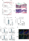
CaMKIV KO mice are protected from experimental colitis. WT and CaMKIV KO mice were subjected to 2% DSS for 7 d. A, B) CaMKIV-deficient mice developed significantly less severe clinical colitis than WT mice, as evidenced by decreased weight loss (A) and decreased DAI scores (B), with a significant difference between WT DSS and CaMKIV KO DSS from d 5 to 7. C) Hematoxylin and eosin staining illustrates in representative images that CaMKIV KO mice displayed significant preservation of intestinal architecture with an intact epithelium and minimal immune cell infiltrate after provocation with 2% DSS vs. WT control mice. Original magnification, ×20. D) Average histology scores, as analyzed by a blinded pathologist. E) CaMKIV-deficient mice demonstrated significantly less FITC-dextran serum absorbance than WT mice. F) Intestinal tissue from WT control and DSS-induced mice and CaMKIV control and DSS-induced mice was analyzed by qPCR for the gene expression of proinflammatory cytokines known to be increased in patients with IBD and mice with induced experimental colitis. G) Levels of oxidized DNA damage was assessed by immunofluorescence with an antibody specific for 8OHdG—a marker for oxidized DNA. CaMKIV KO mice subjected to DSS showed similar levels of oxidized DNA damage compared to WT DSS-induced mice. Images shown are representative images. Scale bar, 50 μm. Values are means ± sd. NS, not significant.
Figure 5
Figure 5
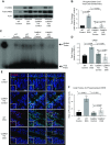
Activation of CaMKIV during intestinal insult induces the activation of CREB. A) Intestinal protein lysates from WT and CaMKIV KO DSS-subjected mice demonstrated an increase in phosphorylated CREB in WT mice vs. CaMKIV KO mice. B) Densitometry illustrates that there was a significant increase in the ratio of phosphorylated/total CREB in WT control vs. WT DSS-induced mice. There is no significant increase in CaMKIV KO mice subjected to DSS. C) EMSAs were performed with oligonucleotides specific for CREB. Incubation of nuclear extracts from DSS-induced WT mice with labeled CREB oligonucleotide resulted in complex formation (as evidenced in the shift of labeled DNA probe). Incubation of nuclear extracts from DSS-induced CaMKIV KO mice with labeled CREB oligonucleotide did not result in complex formation. D) Intestinal tissues were evaluated by qPCR for CREB gene expression. E) Immunofluorescence for phosphorylated CREB (red), E-cadherin (green), and DAPI (blue) was performed in murine intestinal tissue samples. Phosphorylated CREB was present within the intestinal epithelium and was increased in WT mice subjected to DSS. Small solid box on left side of each image is a magnified view of the small dotted boxes. Images shown are a representative image. Scale bar, 50 μm. F) Intensity of phosphorylated CREB immunofluorescence was determined by calculating the CTCF in control and diseased tissue sections. Calculations show an increase in the intensity of phosphorylated CREB in the intestinal epithelium of WT mice subjected to DSS intensity. Values graphed are means ± sd.
Figure 6
Figure 6
Activation of CREB results in cell cycle arrest during intestinal inflammation. A) Intestinal tissues from WT control and DSS-induced mice and CaMKIV control and DSS-induced were evaluated by qPCR for p53 and p21 gene expression. B) Western blot analysis showed an increase in the protein levels of p53 and p21 in WT mice subjected to DSS. CaMKIV KO mice subjected to DSS did not show an increase in the protein levels of p53 and p21. C) Densitometry of immunoblots in B. D) There is a significant decrease in the number of proliferating cells per crypt in WT mice subjected to DSS vs. WT control mice by BrdU analysis. The number of proliferating cells per crypt in CaMKIV KO mice was significantly higher vs. WT DSS-induced mice. Images are representative. E) BrdU+ cells per crypt were counted for each strain (at least 2 crypts per slide and at least 12 different mice). F) Intestinal lysates from control and diseased human tissue samples were analyzed by Western blot for the protein levels of CREB, p53, and p21. G) Densitometry illustrates that there is a significant increase in the phosphorylated:total CREB ratio, p53, and p21 in intestinal tissue from control vs. patients with ulcerative colitis. Values are means ± sd. NS, not significant.
Figure 7
Figure 7
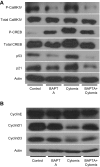
Activation of CaMKIV and subsequent induction of cell cycle arrest in an in vitro model of intestinal inflammation is dependent upon calcium signals. A) Western blot analysis showed that pretreatment of IEC-6 cells exposed to cytomix with BAPTA-AM, a cell-permeant calcium chelator, inhibited activation of CaMKIV and the downstream cell arrest signaling pathway, including p53 and p21. B) Western blot analysis showed that the positive regulators of cell cycle G1-S phase transition cyclin-D1 and -D3 were markedly decreased in IEC-6 cells treated with cytomix, but not in IEC-6 cells treated with BAPTA+cytomix.
Figure 8
Figure 8
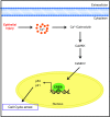
CaMKIV activation plays a critical role in the pathogenesis of IBD. This figure illustrates our current working hypothesis, which outlines a mechanism by which CaMKIV activation leads to intestinal inflammation. Our data support the presence of CaMKIV in IECs and suggest that during intestinal inflammation CaMKIV is activated by a calcium-dependent signal. Activation of CaMKIV induces the activation of the transcription factor CREB, which transactivates p53. p53 upregulates the expression of the proarrest signal p21, which inhibits proliferation of the intestinal epithelium. We hypothesize that this upregulated pathway prevents proliferation of the intestinal epithelium during intestinal inflammation, which ultimately results in a compromised intestinal epithelial barrier and further perpetuates intestinal inflammation.

Similar articles

Cited by

References

    1. Greco C. A., Maurer-Spurej E., Scott M. D., Kalab M., Nakane N., Ramírez-Arcos S. M. (2011) PEGylation prevents bacteria-induced platelet activation and biofilm formation in platelet concentrates. Vox Sang. 100, 336–339 - PubMed
    1. Rigoli L., Caruso R. A. (2014) Inflammatory bowel disease in pediatric and adolescent patients: a biomolecular and histopathological review. World J. Gastroenterol. 20, 10262–10278 - PMC - PubMed
    1. Crohn’s & Colitis Foundation of America. (2014) The Facts about Inflammatory Bowel Disease, Crohn’s & Colitis Foundation of America, New York. Accessed January, 18, 2018, at http://www.crohnscolitisfoundation.org/assets/pdfs/updatedibdfactbook.pdf/
    1. Frolkis A. D., Dykeman J., Negrón M. E., Debruyn J., Jette N., Fiest K. M., Frolkis T., Barkema H. W., Rioux K. P., Panaccione R., Ghosh S., Wiebe S., Kaplan G. G. (2013) Risk of surgery for inflammatory bowel diseases has decreased over time: a systematic review and meta-analysis of population-based studies. Gastroenterology 145, 996–1006 - PubMed
    1. Novak E. A., Mollen K. P. (2015) Mitochondrial dysfunction in inflammatory bowel disease. Front. Cell Dev. Biol. 3, 62 - PMC - PubMed

Publication types

MeSH terms

Substances

LinkOut - more resources