. 2018 Aug 16;13(8):e0201853.
doi: 10.1371/journal.pone.0201853. eCollection 2018.

Generation and characterisation of recombinant FMDV antibodies: Applications for advancing diagnostic and laboratory assays

Affiliations

Generation and characterisation of recombinant FMDV antibodies: Applications for advancing diagnostic and laboratory assays

Gareth Shimmon et al. PLoS One. .

Abstract

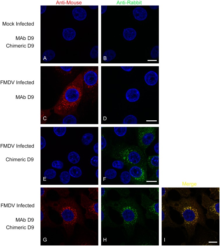
Foot-and-mouth disease (FMD) affects economically important livestock and is one of the most contagious viral diseases. The most commonly used FMD diagnostic assay is a sandwich ELISA. However, the main disadvantage of this ELISA is that it requires anti-FMD virus (FMDV) serotype-specific antibodies raised in small animals. This problem can be, in part, overcome by using anti-FMDV monoclonal antibodies (MAbs) as detecting reagents. However, the long-term use of MAbs may be problematic and they may need to be replaced. Here we have constructed chimeric antibodies (mouse/rabbit D9) and Fabs (fragment antigen-binding) (mouse/cattle D9) using the Fv (fragment variable) regions of a mouse MAb, D9 (MAb D9), which recognises type O FMDV. The mouse/rabbit D9 chimeric antibody retained the FMDV serotype-specificity of MAb D9 and performed well in a FMDV detection ELISA as well as in routine laboratory assays. Cryo-electron microscopy analysis confirmed engagement with antigenic site 1 and peptide competition studies identified the aspartic acid at residue VP1 147 as a novel component of the D9 epitope. This chimeric expression approach is a simple but effective way to preserve valuable FMDV antibodies, and has the potential for unlimited generation of antibodies and antibody fragments in recombinant systems with the concomitant positive impacts on the 3Rs (Replacement, Reduction and Refinement) principles.


Conflict of interest statement

The authors have declared that no competing interests exist.

Figures

Fig 1
Fig 1. Amino acid sequence of the Fv fragment of MAb D9.
The amino acid sequence of the VH (A) and VL (B) domains were aligned using the Chothia and Kabat numbering schemes using the Abnum tool (www.bioinf.org.uk/abs/abnum). The CDR boundaries were assigned using the Chothia (bold) or Kabat (underlined) definitions using the CDR definitions table (www.bioinf.org.uk/abs/#cdrs). Chothia and Kabat numbering is given above and below the sequence alignment respectively. The upper sequence is MAb D9 and the lower MAb SD6.
Fig 2
Fig 2. MAb SD6 and modelling of D9 variable domains.
Cartoon depiction showing SD6 variable domains in grey and modelled D9 VH domain in orange and VL domain in blue. The peptide bound to SD6 is shown in red with the side chains for the RGD motif and the L residues at 144 and 148 drawn in stick representation coloured by atom. R/E indicates the position of the first residue of CDR3, which is an R in SD6 and an E in D9. In the FMDV/SD6 complex the R interacts with VP1 D-147 (interactions are represented by yellow broken lines).
Fig 3
Fig 3. Cryo-EM complex of D9 with FMDV O1Manisa.
The electron potential map of the viral capsid at 4.0 Å resolution is shown as a light blue mesh. Cα traces (shown as continuous thick lines) for the capsid proteins are colour coded: VP1: green, VP2: yellow, VP3: red. The dark blue mesh shows the same map low pass filtered to 20 Å resolution to reveal the mobile Fv portion of D9. For comparison the Cα traces for the variable heavy (purple) and light (green) chains of SD6 complexed with the VP1 GH loop peptide (light blue) are shown as seen in complex with C-serotype virus structure (PDB code: 1qgc), after superimposition of the virus structures. Note that SD6 binds to the VP1 loop at the same point on the capsid but is less upright than suggested for D9 by the diffuse density.
Fig 4
Fig 4. Initial characterisation of the mouse/rabbit D9 chimera.
Panels A and B show a western blot to detect rabbit IgG heavy (A) and light chains (B) in the elution fractions from affinity chromatography of the mouse/rabbit D9 chimera. Panel C shows an ELISA using the elution fractions (E6-12) to detect immobilised FMDV O1K VP1 GH loop peptide. SN = transfected cell culture supernatant; FT = column flow-through; W = column wash; E6-12 = elution fractions 6–12. The OD of the wells was read at 490nm. Shown is the mean and SD of duplicate samples.
Fig 5
Fig 5. Specificity of the mouse/rabbit D9 chimera for type O FMDV.
ELISA 96-well plates were coated with purified ECs (O1Mcc or A22cc), or with coating buffer as indicated on the figure. The ECs were detected using cell supernatants (S) from cells transfected with the expression plasmid for either the HC (S: HCh only), the LC (S: LCh only) or co-transfected with both plasmids (S: HCh & LCh), or with pooled fractions ([fractions 7–12] C: HCh & LCh) of the concentrated mouse/rabbit D9 chimera (shown in Fig 4). To verify capsid immobilisation, the O1Mcc and A22cc ECs were also detected using MAb D9 (Anti-O MAb D9) and a guinea pig, anti-A22Iraq polyclonal sera (Anti-A22 GP) respectively. The OD of the wells was read at 450nm after 0.5h. Shown is the mean and SD of triplicate samples.
Fig 6
Fig 6. A type O FMDV VP1 GH loop peptide inhibits binding of the mouse/rabbit D9 chimera to O1Manisa EC.
ELISA 96-well plates were coated with purified ECs (O1Mcc), or with coating buffer as indicated on the figure. The ECs were detected using the concentrated mouse/rabbit D9 chimera, in the absence or presence of peptides. Peptides: the wt peptide has a sequence (141-VPNLRGDLQVLAQKVAR-158) corresponding to residues 141 to 158 of the VP1 GH loop of type O FMDV (O1K); wt (RGDL) = the wt 17-mer; L148M (RGDM) has a L to M substitution at position 8 of the peptide corresponding to VP1 148; D147E (RGEL) has an D to E substitution at position 7 of the peptide corresponding to VP1 147. The OD of the wells was read at 450nm after 0.5h. Shown is the mean and SD of triplicate samples.
Fig 7
Fig 7. FMDV serotype-specific neutralisation by the mouse/rabbit D9 chimera.
Shown are the virus neutralisation titres for MAb D9 and the concentrated mouse/rabbit D9 chimera (chimeric D9, pooled fractions 7–12). Antibody titres were calculated from regression data as the log 10 reciprocal antibody dilution required for 50% neutralisation of 100 tissue culture infective units of virus (log 10SN 50/100 TCID 50). Shown is the mean and SD of duplicate samples. O1K = FMDV O1 Kaufbeuren; A22 = A22Iraq.
Fig 8
Fig 8. The specificity of the mouse/rabbit D9 chimera for FMDV capsid determined by western blot.
Detection of EC (O1Mcc and A22cc) using MAb D9 (diluted ascites at 1/1000) (A) or the mouse/rabbit D9 chimera (column elution fraction 8 at 1/10) (B).
Fig 9
Fig 9. The mouse/rabbit D9 chimera identifies VP1 in FMDV infected cells.
IBRS-2 cells were mock-treated or infected with type O FMDV. Panels A and B show mock cells incubated with chimeric D9 and MAb D9 simultaneously followed by the goat anti-mouse IgG Alexa-568 and goat anti-rabbit Alexa-488 conjugated secondary antibodies. Panels C and D show infected cells incubated with MAb D9 and both secondary antibodies, and data collection for the red (C) and green (D) channels. Panels E and F show infected cells incubated with the chimeric D9 and both secondary antibodies, and data collection for the red (E) and green (F) channels. Panels G-I show FMDV infected cells incubated simultaneously with chimeric D9 and MAb D9 followed by both secondary antibodies. Panel G shows data collected for MAb D9 and panel H for the chimeric antibody, while panel I shows the merged image (of panels G and H). Panels A and B, C and D, E and F, and G and H, show the same cells imaged using the red or green channels. Scale bar = 10μM.

References

    1. Arzt J, Baxt B, Grubman MJ, Jackson T, Juleff N, Rhyan J, et al. The pathogenesis of foot-and-mouth disease II: viral pathways in swine, small ruminants, and wildlife; myotropism, chronic syndromes, and molecular virus-host interactions. Transboundary and emerging diseases. 2011;58(4):305–26. 10.1111/j.1865-1682.2011.01236.x . - DOI - PubMed
    1. Knight-Jones TJ, Rushton J. The economic impacts of foot and mouth disease—what are they, how big are they and where do they occur? Preventive veterinary medicine. 2013;112(3–4):161–73. 10.1016/j.prevetmed.2013.07.013 . - DOI - PMC - PubMed
    1. Jackson T, King AM, Stuart DI, Fry E. Structure and receptor binding. Virus research. 2003;91(1):33–46. . - PubMed
    1. Rowlands DJ, Cartwright B, Brown F. Evidence for an internal antigen in foot-and-mouth disease virus. The Journal of general virology. 1969;4(4):479–87. 10.1099/0022-1317-4-4-479 . - DOI - PubMed
    1. Talbot P, Brown F. A model for foot-and-mouth disease virus. The Journal of general virology. 1972;15(2):163–9. 10.1099/0022-1317-15-2-163 . - DOI - PubMed

Publication types

MeSH terms

Substances