. 2018 Aug 16;19(1):617.
doi: 10.1186/s12864-018-4995-0.

Discovery and genome characterization of three new Jeilongviruses, a lineage of paramyxoviruses characterized by their unique membrane proteins

Affiliations

Discovery and genome characterization of three new Jeilongviruses, a lineage of paramyxoviruses characterized by their unique membrane proteins

Bert Vanmechelen et al. BMC Genomics. .

Abstract

Background: In the past decade, many new paramyxoviruses that do not belong to any of the seven established genera in the family Paramyxoviridae have been discovered. Amongst them are J-virus (JPV), Beilong virus (BeiPV) and Tailam virus (TlmPV), three paramyxovirus species found in rodents. Based on their similarities, it has been suggested that these viruses should compose a new genus, tentatively called 'Jeilongvirus'.

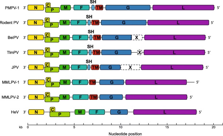
Results: Here we present the complete genomes of three newly discovered paramyxoviruses, one found in a bank vole (Myodes glareolus) from Slovenia and two in a single, co-infected Rungwe brush-furred rat (Lophuromys machangui) from Mozambique, that represent three new, separate species within the putative genus 'Jeilongvirus'. The genome organization of these viruses is similar to other paramyxoviruses, but like JPV, BeiPV and TlmPV, they possess an additional open reading frame, encoding a transmembrane protein, that is located between the F and G genes. As is the case for all Jeilongviruses, the G genes of the viruses described here are unusually large, and their encoded proteins are characterized by a remarkable amino acid composition pattern that is not seen in other paramyxoviruses, but resembles certain motifs found in Orthopneumovirus G proteins.

Conclusions: The phylogenetic clustering of JPV, BeiPV and TlmPV with the viruses described here, as well as their shared features that set them apart from other paramyxoviruses, provide additional support for the recognition of the genus 'Jeilongvirus'.

Keywords: Cell attachment protein; G protein; MMLPV-1; MMLPV-2; PMPV-1; Rodent paramyxovirus.


Conflict of interest statement

Ethics approval and consent to participate

Small mammal sampling in Slovenia was done in cooperation with the Slovenian Museum of Natural History. For the trappings in Mozambique, a small mammal-sampling permit was acquired from the National Directorate for Protected Areas (DINAC – Mozambique). All animal work was approved either by the Administration of the Republic of Slovenia for Food Safety, Veterinary Sector and Plant Protection (U34401–20/2013/5) or by the University of Antwerp Ethical Committee for Animal Experimentation (2011–52).

Consent for publication

Not applicable.

Competing interests

The authors declare that they have no competing interests.

Publisher’s Note

Springer Nature remains neutral with regard to jurisdictional claims in published maps and institutional affiliations.

Figures

Fig. 1
Fig. 1
Nucleotide composition of the PMPV-1 G gene. A sliding window view of the nucleotide composition of the PMPV-1 G gene, using 5% of the gene length as window size, shows that this gene has a low GC-content, comparable to the rest of the PMPV-1 genome, as well as a ~ 1400 nt-long region that is marked by a near-absolute absence of uracils (< 5%) and an abundance of cytosines (up to 50%). The unique nucleotide composition of the PMPV-1 G gene is also reflected in the amino acid composition of the corresponding G protein (see Fig. 5)
Fig. 2
Fig. 2
Maximum clade credibility tree of 69 currently known paramyxovirus species. The phylogeny imputed from the concatenated sequence of the N, P, M, F, G and L proteins of 69 known paramyxovirus species shows that MMLPV-1, MMLPV-2 and PMPV-1 (marked in red) cluster with perfect support as separate species together with JPV, BeiPV, TlmPV and rodent PV. Branches of recognized genera that contain multiple species have been collapsed. An expanded version of this tree is given in Additional file 1: Figure S1. A version of this tree based only on the L protein but including the recently discovered fish paramyxoviruses is given in Additional file 2: Figure S2. Branch lengths are scaled and represent the number of amino acid substitutions per site. Numbers at the different nodes indicate the posterior support for a cluster. GenBank accession numbers of all used sequences are provided in Additional file 3: Table S1
Fig. 3
Fig. 3
Genome organization of all known ‘Jeilongviruses’. All ORFs are drawn to scale. The X ORF (dashed lines) is found only in the genomes of BeiPV, TlmPV and JPV, and is considered part of the G gene. The SH gene is found only in a limited number of paramyxoviruses, while the TM gene appears to be a unique feature of members of the putative genus ‘Jeilongvirus’. Hendra virus (HeV) is included as a reference non-‘Jeilongvirus’ genome
Fig. 4
Fig. 4
Comparison of the G ORFs of all ‘Jeilongviruses’. The G ORFs of the seven known ‘Jeilongviruses’ show a remarkable variation in size, and all of them are larger than those of other paramyxoviruses. The dashed red line marks the average size of a paramyxovirus G ORF. The G ORFs of Tupaia paramyxovirus (TPMV) and Fer-de-lance virus (FDLV) are included to illustrate both the exceptional size of the ‘Jeilongvirus’ G ORFs and their mutual variation, as these two viruses have the largest and smallest G ORFs of all non-‘Jeilongvirus’ paramyxoviruses, respectively. The light blue extensions indicate the complete size of the BeiPV, TlmPV and JPV G genes, including the X ORFs
Fig. 5
Fig. 5
Sliding window overview of the amino acid composition of all Jeilongvirus G proteins. The used window size for each protein is 10% of the respective protein length. The dotted red line represents the average length of paramyxovirus G proteins. All Jeilongvirus G proteins are marked by the presence of an otherwise absent C-terminal region, marked by a high, yet variable abundance of P/T/S (up to 50%). In the genomes of JPV, BeiPV and TlmPV, the G gene is divided into two ORFs, G and X. A single mutation/insertion, however, suffices to join these two ORFs into a single ORF covering the entire G gene. The P/T/S fraction of the resulting hypothetical BeiPV and TlmPV G proteins (yellow/orange dashed lines) is similar to those of the Rodent PV and PMPV-1 G proteins. For JPV, which is more closely related to MMLPV-1/2 (Fig. 2, Additional file 1: Figure S1), this effect is less pronounced, although a narrow peak in P/T/S-abundance, similar to the MMLPV-2 peak, can be observed in the resulting hypothetical JPV G protein (dashed red line)

References

    1. Samal SK. The biology of paramyxoviruses. Norfolk, UK: Caister Academic Press; 2011.
    1. Moss WJ. Measles. Lancet. 2017;390(10111):2490–2502. doi: 10.1016/S0140-6736(17)31463-0. - DOI - PubMed
    1. Rubin S, Eckhaus M, Rennick LJ, Bamford CG, Duprex WP. Molecular biology, pathogenesis and pathology of mumps virus. J Pathol. 2015;235(2):242–252. doi: 10.1002/path.4445. - DOI - PMC - PubMed
    1. Schomacker H, Schaap-Nutt A, Collins PL, Schmidt AC. Pathogenesis of acute respiratory illness caused by human parainfluenza viruses. Curr Opin Virol. 2012;2(3):294–299. doi: 10.1016/j.coviro.2012.02.001. - DOI - PMC - PubMed
    1. Escaffre O, Borisevich V, Rockx B. Pathogenesis of Hendra and Nipah virus infection in humans. J Infect Dev Ctries. 2013;7(4):308–311. doi: 10.3855/jidc.3648. - DOI - PubMed