Comparative Study
. 2018 Aug 16;18(1):28.
doi: 10.1186/s40644-018-0163-7.

Castleman disease versus lymphoma in neck lymph nodes: a comparative study using contrast-enhanced CT

Affiliations
Comparative Study

Castleman disease versus lymphoma in neck lymph nodes: a comparative study using contrast-enhanced CT

Jie Li et al. Cancer Imaging. .

Abstract

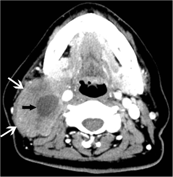
Background: The purpose of this study was to determine the contrast-enhanced CT characteristics for differentiating between Castleman disease (CD) and lymphoma in neck lymph nodes.

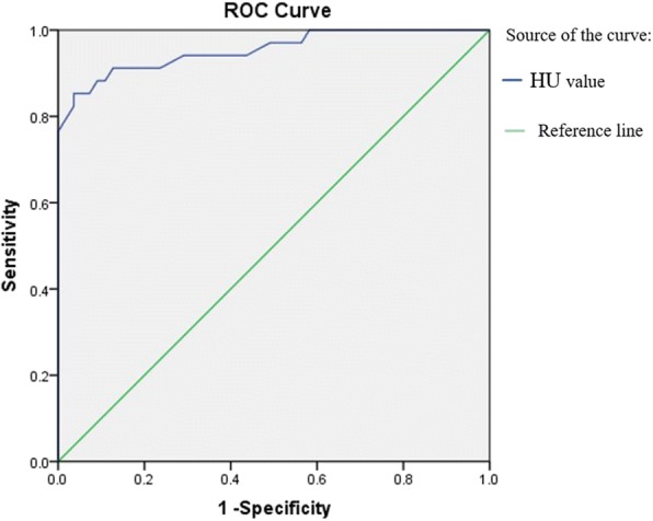
Methods: This retrospective study evaluated the number (solitary or multiple), strength of contrast-enhancement, type of contrast-enhancement, surrounding vessels, contrast-enhanced Hounsfield unit (HU) values, and anatomical distributions of lymph nodes in 34 patients with confirmed CD and 55 patients with newly diagnosed untreated lymphoma. Independent t-tests, receiver operating characteristic (ROC) curve analysis, and chi-square tests were used to evaluate the variables and CT features.

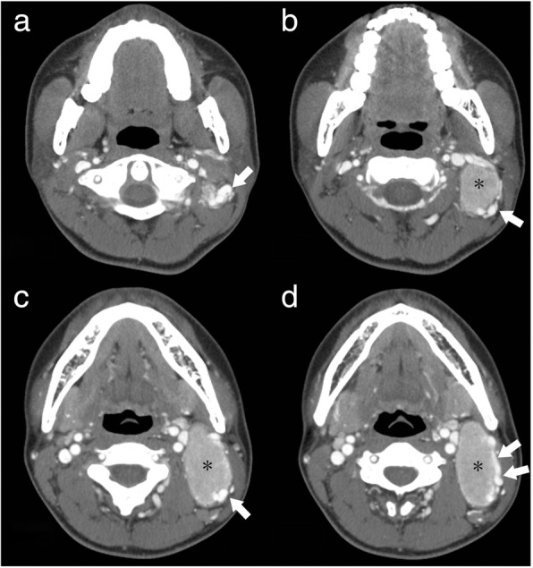
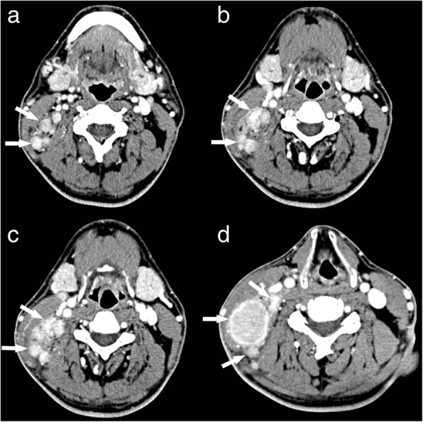
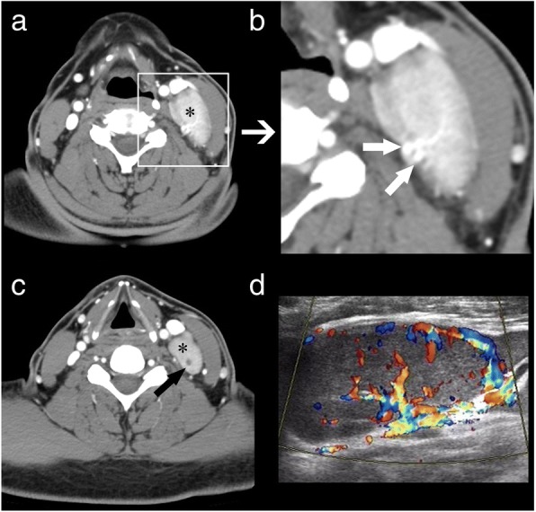
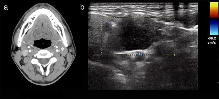
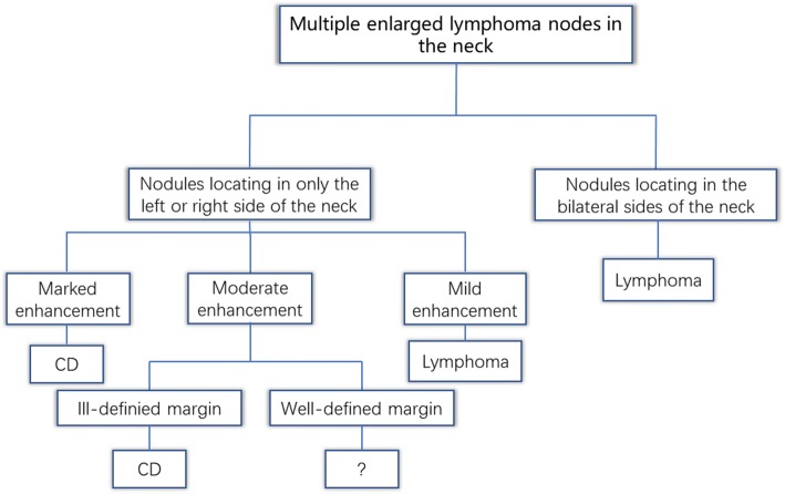
Results: Several significant differences were found between CD and lymphoma. The interval between first contrast-enhanced CT and biopsy/surgery was significantly longer in the CD group (mean 72 ± 105 days, median 60 days) than in the lymphoma patients (mean 30 ± 2 days, median 12 days; p = 0.015). The lymphoma patients presented significantly more often with fatigue and fever (p = 0.023 and p = 0.016 respectively) than did the CD subjects. HU values of nodules after enhancement were significantly higher in the CD patients than in the lymphoma patients. In cases involving multiple lymph nodes, in all the CD cases, all affected nodes were located in only the left or right side of the neck, not bilaterally. ROC analysis showed a significant difference in contrast-enhanced CT attenuation values between lymphoma and CD (p < 0.001, area under the curve = 0.954), with a cut-off value of 92.5 HU. We constructed a decision tree according to these imaging characteristics.

Conclusions: Contrast-enhanced CT can be useful for differentiating between CD and lymphoma.

Keywords: Castleman disease; Contrast-enhanced CT; Lymphoma; Neck.


Conflict of interest statement

Ethics approval and consent to participate

Ethical approval was obtained by the local institutional review board (Ethical Committee of Affiliated Hospital of Qingdao University).

Consent for publication

Not applicable.

Competing interests

The authors declare that they have no competing interests.

Publisher’s Note

Springer Nature remains neutral with regard to jurisdictional claims in published maps and institutional affiliations.

Figures

Fig. 1
Fig. 1
A 29-year-old man who presented with hyaline vascular type Castleman disease. a-d Contrast-enhanced CT images of the neck show a solitary nodule (black *) with a marked enhancement pattern and dilated and tortuous vessels in the periphery of the nodule (white arrows)
Fig. 2
Fig. 2
A 37-year-old man who presented with hyaline vascular type Castleman disease. a-d Contrast-enhanced CT images of the neck show multiple nodules with a marked enhancement pattern (white arrows), with all of the nodules located on the right side of the neck. There are no enlarged lymph nodes on the left side of the neck
Fig. 3
Fig. 3
A 44-year-old man who presented with hyaline vascular type Castleman disease. a, c Contrast-enhanced CT images of the neck show multiple nodules (black *) with marked enhancement patterns. b Dilated and tortuous vessels are observable around the nodules. c An intra-nodular cystic region is also observable (black arrow). d Spectral Doppler ultrasound imaging shows plentiful blood flow
Fig. 4
Fig. 4
A 47-year-old woman who presented with non-Hodgkin’s lymphoma. a A contrast-enhanced CT image of the neck shows a solitary nodule (white *) with a mild enhancement pattern. b Spectral Doppler ultrasound imaging shows no blood flow
Fig. 5
Fig. 5
A 35-year-old man who presented with non-Hodgkin’s lymphoma. a-b Contrast-enhanced CT images of the neck show multiple nodules with mild enhancement patterns (white arrows), with the nodules being located on both sides of the neck (white arrows)
Fig. 6
Fig. 6
A 47-year-old woman who presented with non-Hodgkin’s lymphoma. A contrast-enhanced CT image of the neck shows an ill-defined margin (white arrow) and a region of liquefaction and necrosis with a mild enhancement pattern in the interior of the mass (black arrow)
Fig. 7
Fig. 7
Receiver operating characteristic curves for attenuation values in differentiating CD from lymphoma
Fig. 8
Fig. 8
Decision tree analysis for differentiating CD and lymphoma with multiple lymph node involvement
Fig. 9
Fig. 9
Decision tree analysis for differentiating CD and lymphoma with solitary lymph node involvement

References

    1. Kligerman S, Auerbach A, Franks T, Galvin J. Castleman disease of the thorax: clinical, radiologic, and pathologic correlation: from the radiologic pathology archives. Radiographics. 2016;36(5):1309–1332. doi: 10.1148/rg.2016160076. - DOI - PubMed
    1. Ooi CC, Cheah FK, Wong SK. Castleman's disease of the kidney: sonographic findings. J Clin Ultrasound. 2015;43(7):438–442. doi: 10.1002/jcu.22192. - DOI - PubMed
    1. Ottaviani F, Galli J, Di Girolamo S, Almadori G. Castleman's disease restricted to the parapharyngeal space. J Otolaryngol. 1999;28(2):95–98. - PubMed
    1. Bonekamp D, Hruban RH, Fishman EK. The great mimickers: Castleman disease. Semin Ultrasound CT MR. 2014;35(3):263–271. doi: 10.1053/j.sult.2013.12.005. - DOI - PubMed
    1. Jiang XH, Song HM, Liu QY, Cao Y, Li GH, Zhang WD. Castleman disease of the neck: CT and MR imaging findings. Eur J Radiol. 2014;83(11):2041–2050. doi: 10.1016/j.ejrad.2014.08.013. - DOI - PubMed

Publication types