. 2018 Sep 28;293(39):15208-15220.
doi: 10.1074/jbc.RA118.003831. Epub 2018 Aug 16.

Mechanism of dysfunction of human variants of the IRAK4 kinase and a role for its kinase activity in interleukin-1 receptor signaling

Affiliations

Mechanism of dysfunction of human variants of the IRAK4 kinase and a role for its kinase activity in interleukin-1 receptor signaling

Saurav De et al. J Biol Chem. .

Abstract

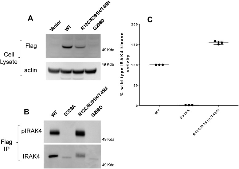
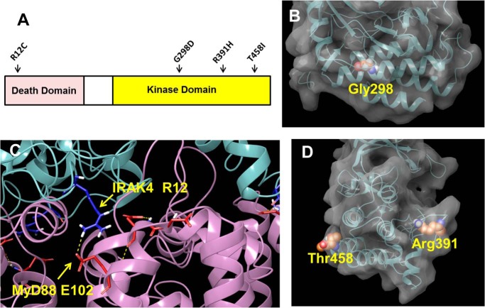
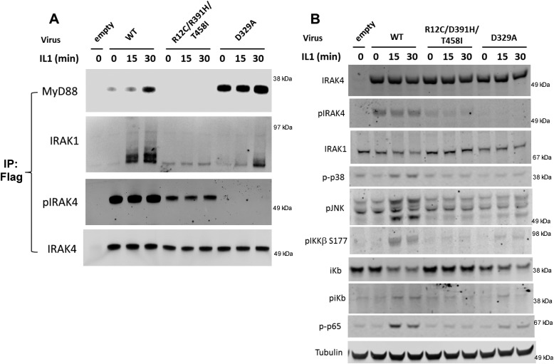
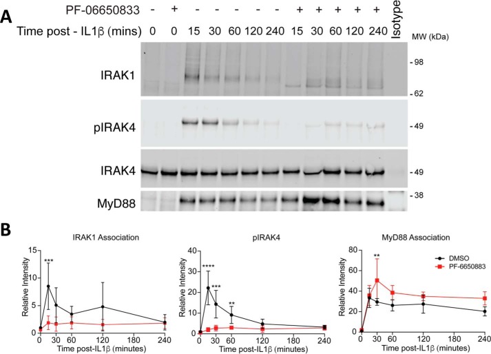
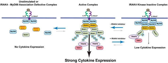
Interleukin-1 receptor (IL1R)-associated kinase 4 (IRAK4) is a central regulator of innate immune signaling, controlling IL1R and Toll-like receptor (TLR)-mediated responses and containing both scaffolding and kinase activities. Humans deficient in IRAK4 activity have autosomal recessive primary immune deficiency (PID). Here, we characterized the molecular mechanism of dysfunction of two IRAK4 PID variants, G298D and the compound variant R12C (R12C/R391H/T458I). Using these variants and the kinase-inactive D329A variant to delineate the contributions of IRAK4's scaffolding and kinase activities to IL1R signaling, we found that the G298D variant is kinase-inactive and expressed at extremely low levels, acting functionally as a null mutation. The R12C compound variant possessed WT kinase activity, but could not interact with myeloid differentiation primary response 88 (MyD88) and IRAK1, causing impairment of IL-1-induced signaling and cytokine production. Quantitation of IL-1 signaling in IRAK4-deficient cells complemented with either WT or the R12C or D329A variant indicated that the loss of MyD88 interaction had a greater impact on IL-1-induced signaling and cytokine expression than the loss of IRAK4 kinase activity. Importantly, kinase-inactive IRAK4 exhibited a greater association with MyD88 and a weaker association with IRAK1 in IRAK4-deficient cells expressing kinase-inactive IRAK4 and in primary cells treated with a selective IRAK4 inhibitor. Loss of IRAK4 kinase activity only partially inhibited IL-1-induced cytokine and NF-κB signaling. Therefore, the IRAK4-MyD88 scaffolding function is essential for IL-1 signaling, but IRAK4 kinase activity can control IL-1 signal strength by modulating the association of IRAK4, MyD88, and IRAK1.

Keywords: Toll/interleukin-1 receptor (TIR); cell signaling; cytokine; inflammation; innate immunity; interleukin 1 (IL-1); interleukin-1 receptor-associated kinase 4 (IRAK4); myeloid differentiation primary response gene 88 (MyD88); protein kinase.


Conflict of interest statement

S. D., F. K,. and V. R. R. are paid employees of Pfizer Inc. E. K., L. C., and L.-L. L. were paid employees of Pfizer Inc. at the time of this work

Figures

Figure 1.
Figure 1.
Characterization of IRAK4 variant patient dermal fibroblasts. SV40-transformed human dermal fibroblasts were stimulated with 10 ng/ml IL-1β and assayed for phosphoprotein expression and cytokines (WT homozygote (WT); IRAK4-null homozygote (P15), and R12C.R391H.T458I/IRAK4-null heterozygote (R12C)). A, immunoblots of whole-cell lysates at the indicated times of IL-1β stimulation. The blot is representative of three independent experiments. B, quantitation of IRAK4 expression as determined by immunoblot in A and normalized to tubulin. C, measurement of secreted IL-8 following 4 h of IL-1β treatment. Data reflect the mean of three independent experiments, significance determined by one-way analysis of variance. NS, not significant. Error bars, S.D.
Figure 2.
Figure 2.
Expression and kinase activity of variants of IRAK4. A, Western blotting of cell lysates from COS7 cells overexpressing IRAK4 constructs. B, Western blotting of FLAG immunoprecipitated autophosphorylated IRAK4 constructs expressed in COS7 cells. C, kinase activity of purified IRAK4 constructs expressed in COS7 cells normalized to IRAK4 protein expression. Data reflect the mean of three independent experiments. IP, immunoprecipitation. Error bars, S.E.
Figure 3.
Figure 3.
Position of mutations in IRAK4 structure. A, cartoon showing position of human mutations in IRAK4. B, space-fill representation of Gly-298 in the kinase domain of IRAK4 taken from PDB entry 2NRU. C, interaction of IRAK4 Arg-12 with Glu-102 of MyD88 taken from PDB entry 3MOP. D, space-fill representations of Thr-458 and Arg-391 in the kinase domain of IRAK4 taken from PDB entry 2NRU. Space-fill representations were created using the Maestro software program (Schrödinger LLC, New York).
Figure 4.
Figure 4.
IL-1–induced IL-8 production in IRAK4-null dermal fibroblasts reconstituted with IRAK4 variants. A, reconstitution of IL-1–induced IL-8 cytokine production. Human IRAK4-null dermal fibroblasts (P15 from Fig. 1A) were infected with the indicated MOIs of WT, R12C/R391H/T458I, and D329A IRAK4 adenovirus for 72 h and stimulated for 4 h with 10 ng/ml IL-1β. B, Western blotting of IRAK4 and tubulin from A. C, IL-8 levels measured in A normalized to IRAK4 expression levels determined in B and expressed as a percentage of WT activity. Data reflect the mean from four independent experiments. Error bars, S.D.
Figure 5.
Figure 5.
Reconstitution of IRAK4-deficient dermal fibroblasts with IRAK4 mutations. Adenovirus containing WT, R12C/R391H/T458I, or D329A FLAG-tagged constructs of IRAK4 was transduced into IRAK4-deficient dermal fibroblasts for 72 h and stimulated with 10 ng/ml IL-1β for the indicated time. A, FLAG immunoprecipitation of cells followed by Western blotting with the indicated antibodies. B, whole-cell lysates from A probed with phosphospecific antibodies against the indicated proteins. Images represent one example of three independent experiments. IP, immunoprecipitation.
Figure 6.
Figure 6.
Inhibition of IL-1–induced signaling and cytokine in primary dermal fibroblasts using an IRAK4 inhibitor. A, primary human neonatal dermal fibroblasts in the presence of either DMSO or 200 nm IRAK4 inhibitor PF-06650833 were stimulated with 2 ng/ml IL-1β overnight, after which IL-6 and IL-8 secretion was measured. B, IL-6 and IL-8 transcripts as determined following stimulation with 2 ng/ml IL-1β for 6 h in the presence of either DMSO or 200 nm IRAK4 inhibitor PF-06650833. C, primary human neonatal dermal fibroblasts were stimulated with 2 ng/ml IL-1β for the indicated times in the presence of DMSO or 200 nm PF-06650833 and lysed and immunoblotted for the indicated proteins. D, quantitation of phosphoproteins from C relative to parent protein (DMSO (black) or PF-06650833 (red)). Data represent the mean of three independent experiments.
Figure 7.
Figure 7.
Immunoprecipitation of IRAK4-associated proteins from primary human dermal fibroblasts in the presence of the selective IRAK4 inhibitor PF-06650833. A, primary human neonatal dermal fibroblasts stimulated with 2 ng/ml IL-1β for the indicated time in the presence or absence of 200 nm PF-06650833 were lysed, and IRAK4 was immunoprecipitated and immunoblotted for the indicated proteins. The image represents one example of four independent experiments. B, quantitation of proteins from A relative to total IRAK4 (DMSO (black) or PF-06650833 (red)). Data represent the mean of four independent experiments. Error bars, S.D.
Figure 8.
Figure 8.
Scheme showing the role of kinase and scaffolding activity of IRAK4 in IL-1 signaling. In the unstimulated complex or the MyD88 interaction–incompetent R12C IRAK4 variant (left), no myddosome is formed, leading to complete loss of IL-1 signaling. In the active complex (center) a myddosome is formed with weak association of IRAK4 and MyD88 and strong association of IRAK4 and IRAK1 that potentiates robust ubiquitination and phosphorylation of IRAK1 and strong downstream signaling from the IL-1 receptor. In the IRAK4 kinase–inhibited complex (right), a myddosome is formed where IRAK4 is strongly associated with MyD88 but weakly associated with IRAK1, which reduces ubiquitination of IRAK1 and decreases IL-1–induced signaling and cytokine production.

References

    1. Alsina L., Israelsson E., Altman M. C., Dang K. K., Ghandil P., Israel L., von Bernuth H., Baldwin N., Qin H., Jin Z., Banchereau R., Anguiano E., Ionan A., Abel L., Puel A., et al. (2014) A narrow repertoire of transcriptional modules responsive to pyogenic bacteria is impaired in patients carrying loss-of-function mutations in MYD88 or IRAK4. Nat. Immunol. 15, 1134–1142 10.1038/ni.3028 - DOI - PMC - PubMed
    1. Dossang A. C., Motshwene P. G., Yang Y., Symmons M. F., Bryant C. E., Borman S., George J., Weber A. N., and Gay N. J. (2016) The N-terminal loop of IRAK-4 death domain regulates ordered assembly of the myddosome signalling scaffold. Sci. Rep. 6, 37267 10.1038/srep37267 - DOI - PMC - PubMed
    1. Israel L., Wang Y., Bulek K., Della Mina E., Zhang Z., Pedergnana V., Chrabieh M., Lemmens N. A., Sancho-Shimizu V., Descatoire M., Lasseau T., Israelsson E., Lorenzo L., Yun L., Belkadi A., et al. (2017) Human adaptive immunity rescues an inborn error of innate immunity. Cell 168, 789–800.e10 10.1016/j.cell.2017.01.039 - DOI - PMC - PubMed
    1. Picard C., Casanova J. L., and Puel A. (2011) Infectious diseases in patients with IRAK-4, MyD88, NEMO, or IκBα deficiency. Clin. Microbiol. Rev. 24, 490–497 10.1128/CMR.00001-11 - DOI - PMC - PubMed
    1. Vollmer S., Strickson S., Zhang T., Gray N., Lee K. L., Rao V. R., and Cohen P. (2017) The mechanism of activation of IRAK1 and IRAK4 by interleukin-1 and Toll-like receptor agonists. Biochem. J. 474, 2027–2038 10.1042/BCJ20170097 - DOI - PMC - PubMed

Publication types

MeSH terms

LinkOut - more resources