. 2018 Sep 28;361(6409):1386-1389.
doi: 10.1126/science.aat8849. Epub 2018 Aug 16.

A mechanism for preventing asymmetric histone segregation onto replicating DNA strands

Affiliations

A mechanism for preventing asymmetric histone segregation onto replicating DNA strands

Chuanhe Yu et al. Science. .

Abstract

How parental histone (H3-H4)2 tetramers, the primary carriers of epigenetic modifications, are transferred onto leading and lagging strands of DNA replication forks for epigenetic inheritance remains elusive. Here we show that parental (H3-H4)2 tetramers are assembled into nucleosomes onto both leading and lagging strands, with a slight preference for lagging strands. The lagging-strand preference increases markedly in budding yeast cells lacking Dpb3 and Dpb4, two subunits of the leading strand DNA polymerase, Pol ε, owing to the impairment of parental (H3-H4)2 transfer to leading strands. Dpb3-Dpb4 binds H3-H4 in vitro and participates in the inheritance of heterochromatin. These results indicate that different proteins facilitate the transfer of parental (H3-H4)2 onto leading versus lagging strands and that Dbp3-Dpb4 plays an important role in this poorly understood process.


Figures

Fig. 1.
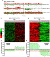
Fig. 1.. Newly synthesized (H3K56ac) and parental histone (H3K4me3) show a slight preference for leading and lagging strands, respectively.
(A–B) An outline of experimental procedures (A) and a diagram for the hypothetic eSPAN outcome assuming four parental and four new (H3–H4)2 tetramers are equally distributed to leading and lagging strand (B). (C) Heatmap showing the bias pattern of H3K56ac eSPAN peaks at each of the 20 individual nucleosomes surrounding 134 early DNA replication origins. The individual nucleosome position is numbered from (−10 to +10) and represented by a circle (top panel). Each row represents the average log2 ratio Watson/Crick H3K56ac eSPAN sequence reads at one of the 134 origins and is clustered based on hierarchical clustering analysis. (D) The average bias ratio of H3K56ac eSPAN peaks at each of the 20 individual nucleosomes of the 134 early replication origins from three independent experiments. (E–F) H3K4me3 eSPAN peaks at newly replicated chromatin exhibit a slight lagging strand bias.
Fig. 2.
Fig. 2.. Analyzing nucleosome assembly of new and parental histone H3 in dpb3Δ cells using the Recom bination-Induced Tag Exchange (RITE) system.
(A) Schematic outline for marking the parental and newly synthesized H3 with the HA epitope (H3-HA) and T7 (H3-T4), respectively. (B) Heat map showing H3-T7 eSPAN bias pattern in dpb3Δ cells at each of 134 individual origins ranked from top to bottom based on the replication efficiency. (C) The average bias pattern of H3-T7 eSPAN peaks at 134 early replication origins. (D–E) H3-HA eSPAN peaks in dpb3Δ cells show a strong lagging strand bias.
Fig. 3.
Fig. 3.. Deletion of DPB3 results in a marked increase in the bias ratio of H3K56ac and H3K4me3 eSPAN peaks under norm al cell cycle progression.
(A) A snapshot of BrdU IP-ssSeq, H3K56ac and H3K4me3 eSPAN peaks at individual nucleosomes surrounding ARS1309 in dpb3Δ cells. (B–D) Analysis of the average bias pattern of H3K56ac eSPAN peaks (B–C) and H3K4me3 eSPAN peaks (D–E) in dpb3Δ cells at normal S phase (n=2). H3K56ac and H3K4me3 eSPAN were performed using dpb3Δ cells released from the G1 block into fresh media without HU at 25°C for 30 min.
Fig. 4.
Fig. 4.. Deletion of DPB3 and DPB4 compromises the parental (H3–H4)2 transfer to leading strands.
(A) A diagram for calculating the relative amount of histone (H3–H4)2 at leading and lagging strand in dpb3A cells compared to wild type using the eSPAN datasets. Grey circles: parental (H3–H4)2; orange circles: new (H3–H4)2; Black line: parental DNA; Red and Green lines: newly synthesized Watson and Crick strand, respectively. (B) Analysis of the relative levels of H3K4me3 in dpb3Δcells compared to wild type at each of the 134 individual origins. 0: no difference between mutant and wild type (black); 003C0 and >0 represent less (green color) and more (red color) H3K4me3 eSPAN sequence reads in dpb3Δ cells than in wild type cells, respectively. Bottom panel: the average amount of H3K4me3 at lagging and leading strands of 134 replication origins in dpb3Δ cells over wild type. (C) Dpb3 and Dpb4 eSPAN peaks at the ARS1309 origin. (D) The average bias of Dpb3 and Dpb4 eSPAN peaks at 134 early replication origins. (E) GST-Dpb3-Dpb4 pull down assays show that Dpb3-Dpb4 dimers bind recombinant (H3–H4)2 tetramers in vitro. (F) dpb3Δ and dpb4Δcells exhibit an increased loss of silencing at the HML locus. (G) A role for Dpb3-Dpb4 in the transfer of parental histone (H3–H4)2 onto leading strands.

Comment in

  • No strand left behind.
    Ahmad K, Henikoff S. Ahmad K, et al. Science. 2018 Sep 28;361(6409):1311-1312. doi: 10.1126/science.aav0871. Science. 2018. PMID: 30262484 No abstract available.

References

    1. Coleman RT, Struhl G, Science 356, 41–51 (2017). - PMC - PubMed
    1. Ragunathan K, Jih G, Moazed D, Science 348, 1258699 (2015). - PMC - PubMed
    1. Laprell F, Finkl K, Muller J, Science 356, 85–88 (2017). - PubMed
    1. Wang X, Moazed D, Science 356, 88–91 (2017). - PMC - PubMed
    1. Campos EI, Stafford JM, Reinberg D, Trends Cell Biol 24, 664–674 (2014). - PMC - PubMed

Publication types