. 2018 Aug 16;10(8):2016-2036.
doi: 10.18632/aging.101526.

Lycopene ameliorates oxidative stress in the aging chicken ovary via activation of Nrf2/HO-1 pathway

Affiliations

Lycopene ameliorates oxidative stress in the aging chicken ovary via activation of Nrf2/HO-1 pathway

Xingting Liu et al. Aging (Albany NY). .

Abstract

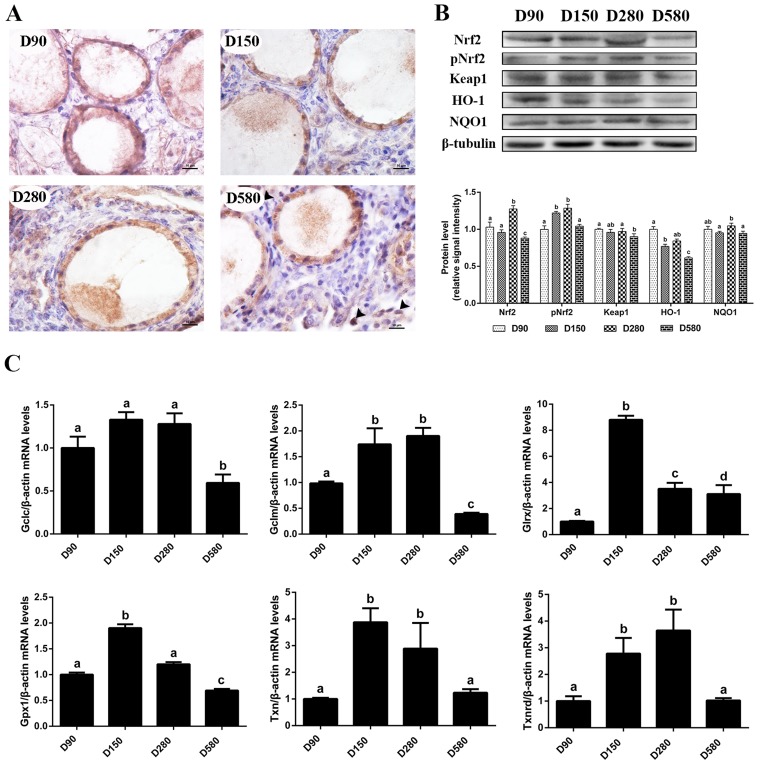
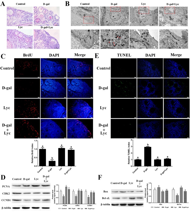
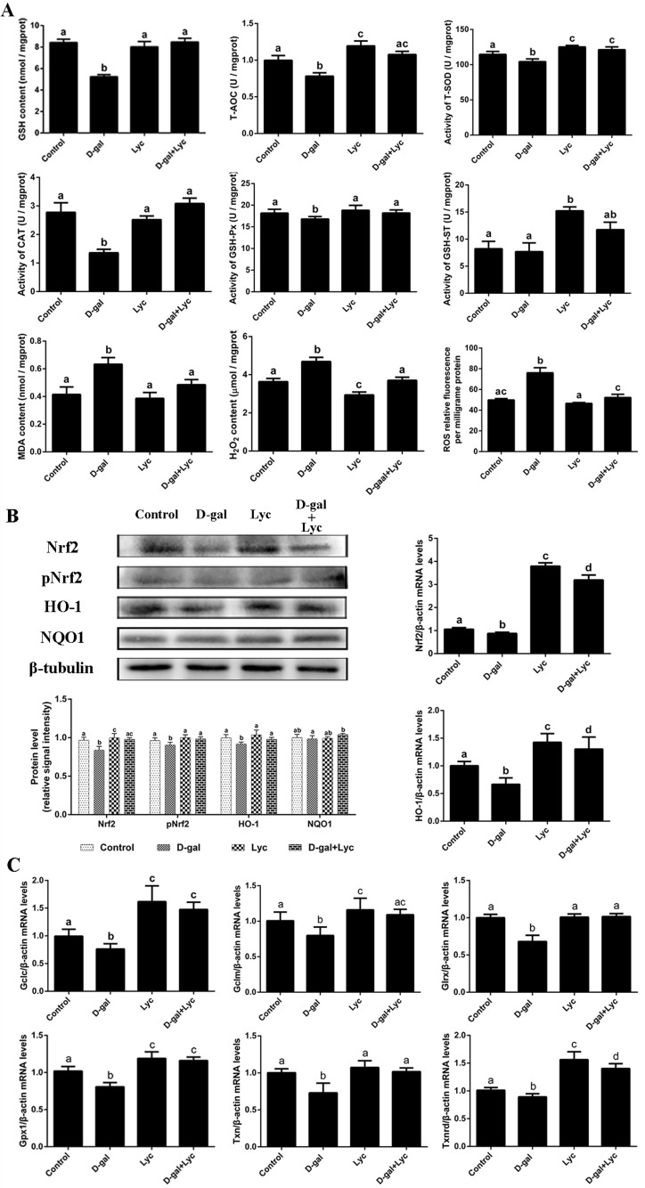
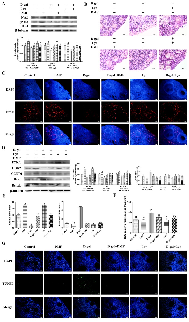
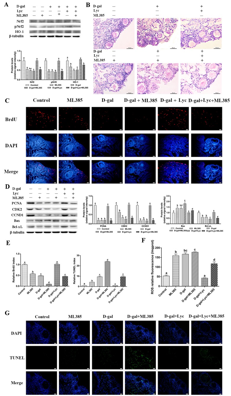
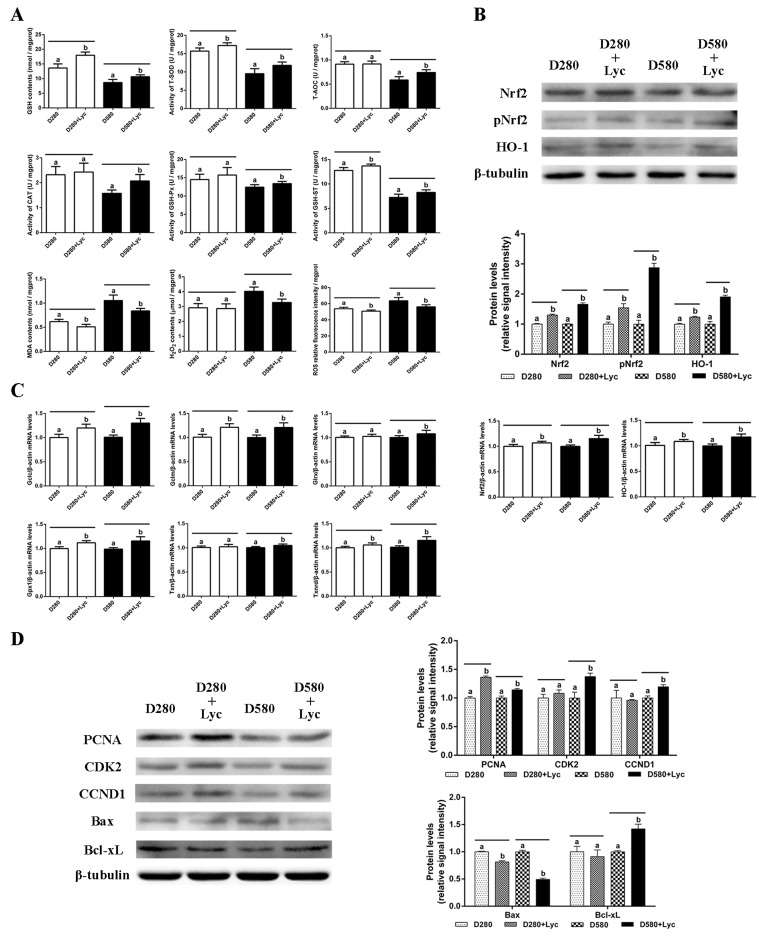
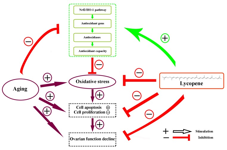
After 480 days of age, high-producing hens are likely to be subject to ovarian aging, mainly due to oxidative stress. In this study, the amelioration of ovarian aging in chickens, using a plant antioxidant, lycopene, was investigated. The activity of the Nrf2/HO-1 pathway in chicken ovaries at different ages (90, 150, 280 and 580 days old) were compared to elucidate any age-related changes. Subsequently, the putative attenuating effect of lycopene (100 ng/mL) on ovarian aging was evaluated through the establishment of a D-gal-induced aging ovarian culture model. The cultured ovarian tissues of young (280 days) and old (580 days) hens were treated with lycopene for 72 h to verify protective effects of lycopene on naturally aged ovaries. Results showed that the Nrf2/HO-1 pathway was down-regulated during the ovarian aging process. Lycopene rescued the decreased antioxidant capacity by increasing the activities of antioxidases and activating the Nrf2/HO-1 pathway in both D-gal-induced and naturally aged ovaries. Moreover, lycopene promoted cell proliferation and inhibited apoptosis in both D-gal-induced and naturally aged ovaries. Lycopene also alleviated D-gal-induced mitochondrial damage in the living granulosa cells. In conclusion, lycopene can effectively ameliorate the oxidative stress in aging hen ovaries via the activation of the Nrf2/HO-1 pathway.

Keywords: Nrf2/HO-1; chicken; lycopene,ovarian aging; oxidative stress.


Conflict of interest statement

CONFLICTS OF INTEREST: All authors declare that they have no conflict of interests.

Figures

Figure 1
Figure 1
Age-related changes in the activity of the Nrf2/HO-1 pathway. (A) Immunohistochemistry of Nrf2 in the ovaries of hens aged 90, 150, 280 and 580 days, scale bar: 10 μm, black arrowheads: Nrf2 located in the nucleus. (B) Age-related changes in relative expression levels of Nrf2, pNrf2, Keap1, HO-1 and NQO1. (C) Age-related changes in transcription levels of Nrf2/HO-1 downstream genes: Gclc, Gclm, Glrx, Gpx1, Txn, Txnrd. Values are expressed as the means±s.e.. The relative abundance of each transcript was normalized to a β-actin and expressed as fold change over D90 ovaries. Different lowercase letters indicate significant differences (P < 0.05).
Figure 2
Figure 2
Protective effects of lycopene on D-gal-induced aged ovaries. (A) Effect of lycopene on D-gal-induced morphological changes of ovarian tissues, scale bar: 50 µm. (B) Effect of lycopene on D-gal-induced ultrastructural changes of granulosa cells: the four pictures in the lower row are the higher magnifications of the red squares from the four pictures in the upper row, respectively, scale bar: 2 µm (upper); 1 µm (lower), red arrowheads: fragmented mitochondria. (C) Effect of lycopene on D-gal-induced decline of BrdU index, scale bar: 20 µm; (D) Relative expression of proteins related to cell proliferation. (E) Effect of lycopene on D-gal-induced increase of TUNEL index, scale bar: 20 µm. (F) Relative expression of proteins related to pro-apoptosis (Bax) and anti-apoptosis (Bcl-xL). Values are expressed as the means±s.e.. Different lowercase letters indicate significant differences (P < 0.05).
Figure 3
Figure 3
Effects of lycopene on decreased antioxidant status in D-gal-induced aged ovarian tissues and the activities of Nrf2/HO-1 pathway. (A) Effect of lycopene on decreased antioxidants status in the D-gal-induced aged ovarian tissues. (B) Effect of lycopene on the down-regulated expression of Nrf2, pNrf2 and HO-1, and the mRNA abundance of Nrf2 and HO-1. (C) Effect of lycopene on down-regulated mRNA abundance of Nrf2/HO-1 downstream genes. Values are expressed as the means±s.e.. Different lowercase letters indicate significant differences (P < 0.05).
Figure 4
Figure 4
Protective effect of lycopene on oxidative stress in aged ovarian tissues was similar to the effects of DMF. (A) Relative changes in the expression of Nrf2, pNrf2 and HO-1 after treatment with D-gal alone or combined with DMF or lycopene. (B) Changes in the morphology of ovarian tissues after treatment with D-gal alone or combined with DMF or lycopene, scale bar: 50 µm. (C and E) Changes in BrdU index in ovarian tissues after treatment with D-gal alone or combined with DMF or lycopene, scale bar: 20 µm. (D) Relative changes in the expression of proteins related to cell proliferation and cell apoptosis in ovarian tissues after treatment with D-gal alone or combined with DMF or lycopene. (E and G) Changes in TUNEL index in ovarian tissues after treatment with D-gal alone or combined with DMF or lycopene, scale bar: 20 µm. (F) Changes in ROS levels of the ovarian tissues after treatment with D-gal alone or combined with DMF or lycopene. Values are expressed as the means±s.e.. Different lowercase letters indicate significant differences (P < 0.05).
Figure 5
Figure 5
Protective effect of lycopene on the oxidative stress in the aging ovarian tissues was abolished by ML385. (A) Relative changes in the expression of Nrf2, pNrf2 and HO-1 after treatment with D-gal, ML385 alone or combined with lycopene. (B) Changes in the morphology of ovarian tissues after treatment with D-gal, ML385 alone or combined with lycopene, scale bar: 50 µm. (C and E) Changes in BrdU index in ovarian tissues after treatment with D-gal, ML385 alone or combined with lycopene, scale bar: 20 µm. (D) Relative changes in the expression of proteins related to cell proliferation and cell apoptosis in ovarian tissues after treatment with D-gal, ML385 alone or combined with lycopene. (E and G) Changes in TUNEL index in ovarian tissues after treatment with D-gal, ML385 alone or combined with lycopene, scale bar: 20 µm. (F) Changes in the levels of ROS in ovarian tissues after treatment with D-gal, ML385 alone or combined with lycopene. Values are expressed as the means±s.e.. Different lowercase letters indicate significant differences (P < 0.05).
Figure 6
Figure 6
Effects of lycopene on the antioxidant capacity, cell proliferation and apoptosis in the ovarian tissues of D280 and D580 hens in vitro. (A) Effect of lycopene on antioxidant capacity in ovarian tissues of D280 and D580 hens in vitro. (B) Effect of lycopene on expression of Nrf2, pNrf2 and HO-1, and the mRNA abundance of Nrf2 and HO-1 in ovarian tissues of D280 and D580 hens in vitro. (C) Effect of lycopene on the mRNA abundance of Nrf2/HO-1 downstream genes in the ovarian tissues of D280 and D580 hens in vitro. (D) Effect of lycopene on the expression of proteins related to cell proliferation and apoptosis in the ovarian tissues of D280 and D580 hens in vitro. Each parameter was determined after 72 h of treatment with lycopene (100 ng/mL). Values are expressed as the means±s.e.. Different lowercase letters indicate significant differences (P < 0.05) for the same age.
Figure 7
Figure 7
Schematic diagram summarizing the mechanisms underlying the attenuating effect of lycopene against ovarian oxidative stress during the aging process in chickens. The Nrf2/HO-1 pathway was down-regulated in the natural aging process in the laying hens. Lycopene attenuated the oxidative stress in aging ovaries via the activation of Nrf2/HO-1 pathway.

References

    1. Mol BW, Zoll M. Fertility preservation for age-related fertility decline. Lancet. 2015; 385:507. 10.1016/S0140-6736(15)60199-4 - DOI - PubMed
    1. Perheentupa A, Huhtaniemi I. Aging of the human ovary and testis. Mol Cell Endocrinol. 2009; 299:2–13. 10.1016/j.mce.2008.11.004 - DOI - PubMed
    1. Ben-Meir A, Burstein E, Borrego-Alvarez A, Chong J, Wong E, Yavorska T, Naranian T, Chi M, Wang Y, Bentov Y, Alexis J, Meriano J, Sung HK, et al.. Coenzyme Q10 restores oocyte mitochondrial function and fertility during reproductive aging. Aging Cell. 2015; 14:887–95. 10.1111/acel.12368 - DOI - PMC - PubMed
    1. Chiang T, Schultz RM, Lampson MA. Age-dependent susceptibility of chromosome cohesion to premature separase activation in mouse oocytes. Biol Reprod. 2011; 85:1279–83. 10.1095/biolreprod.111.094094 - DOI - PMC - PubMed
    1. Herbert M, Kalleas D, Cooney D, Lamb M, Lister L. Meiosis and maternal aging: insights from aneuploid oocytes and trisomy births. Cold Spring Harb Perspect Biol. 2015; 7:a017970. 10.1101/cshperspect.a017970 - DOI - PMC - PubMed