. 2018 Aug 16;19(8):2421.
doi: 10.3390/ijms19082421.

Intrathecal Injection of Dual Zipper Kinase shRNA Alleviating the Neuropathic Pain in a Chronic Constrictive Nerve Injury Model

Affiliations

Intrathecal Injection of Dual Zipper Kinase shRNA Alleviating the Neuropathic Pain in a Chronic Constrictive Nerve Injury Model

Meei-Ling Sheu et al. Int J Mol Sci. .

Abstract

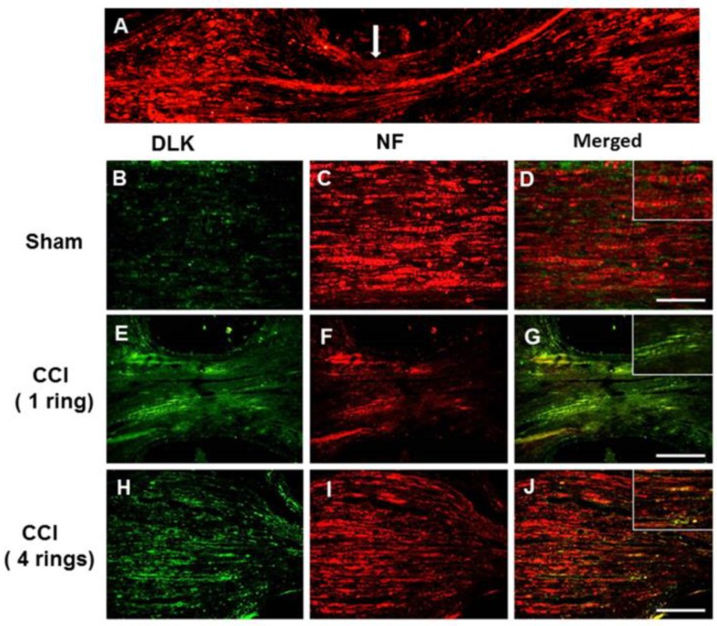
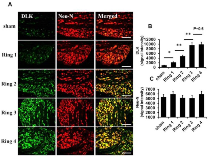
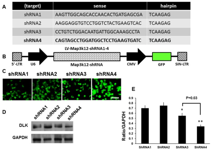
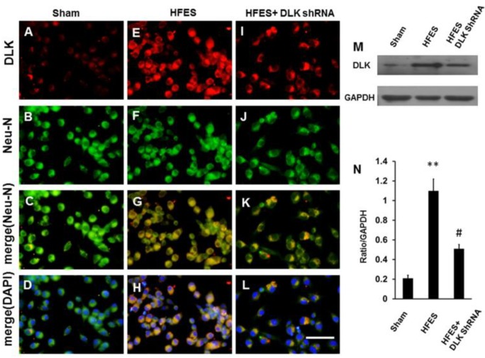
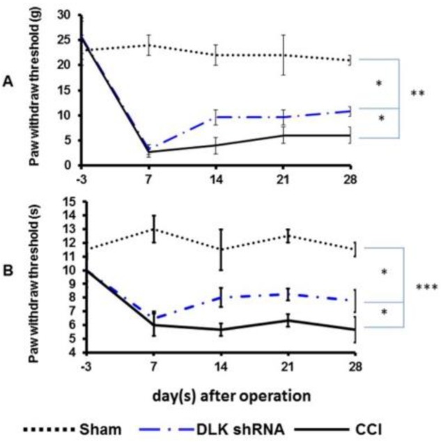
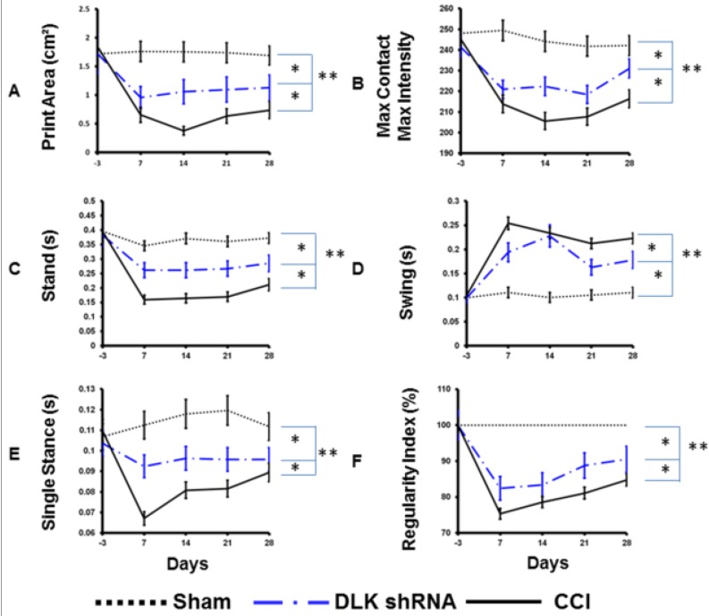
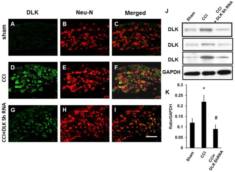
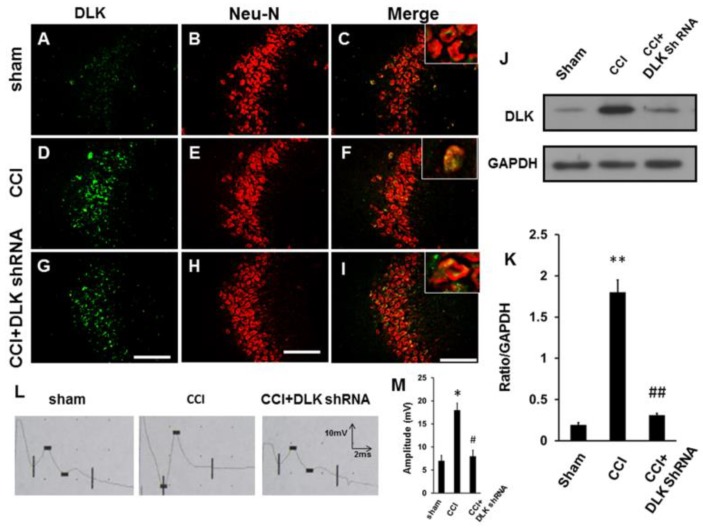
Dual leucine zipper kinase (DLK) is a member of mitogen-activated protein kinase kinase kinase (MAP3K) family mainly involved in neuronal degeneration. However, the role of DLK signaling in the neuropathic pain has not yet been fully determined. Chronic constrictive injury (CCI) was conducted by four 3-0 chromic gut ligatures loosely ligated around the sciatic nerve. Escalated DLK expression over the dorsal root ganglion was observed from one to four rings of CCI. Remarkable expression of DLK was observed in primary dorsal root ganglion cells culture subjected to electrical stimulation and attenuated by DLK short hairpin RNA (shRNA) treatment. Intrathecal injection of DLK shRNA attenuates the expression of DLK over the dorsal root ganglion and hippocampus neurons and increased the threshold of mechanical allodynia and decreased thermal hyperalgesia. In CatWalk gait analysis, significant decreases of print area, maximum contact maximum intensity, stand phase, single stance, and regular index by CCI were alleviated by the DLK shRNA administration. In conclusion, the expression of DLK was up-regulated in chronic constrictive injury and attenuated by the administration of DLK shRNA, which paralleled the improvement of neurobehavior of neuropathic pain. The modulation of DLK expression is a potential clinic treatment option for neuropathic pain.

Keywords: chronic constrictive injury; dual zipper kinase; neuropathic pain.


Conflict of interest statement

The authors declare no conflict of interest.

Figures

Figure 1
Figure 1
Representative dual leucine zipper kinase (DLK) and neurofilament (NF) expression over the injury site of the nerve in chronic constrictive injury (CCI) animals four weeks after injury. (A) Distribution of DLK distal and proximal to the point of CCI injury. (B) Distribution of DLK in the proximal site of the sham group. (C) Co-localized with Neurofilament. (D) Merged imaging fusion of (B,C), right upper box indicated the imaging amplification. (E) Distribution of DLK in the proximal site of one ring CCI. (F) Co-localized with Neurofilament. (G) Merged imaging fusion of E and F, right upper box indicated imaging amplification. (H) Distribution of DLK in the proximal site of four rings CCI. (I) Co-localized with Neurofilament. (J) Merged imaging fusion of (H,I), right upper box indicated imaging amplification. Bar length = 200 μm; Arrow indicated the nerve crushed region.
Figure 2
Figure 2
Depiction of DLK expression over the dorsal root ganglia cells subjected to the escalating numbers of ligature rings from one to four in animals four weeks after CCI. (A) Illustration of DLK expression over the dorsal root ganglion cells co-localized with Neu-N related to the escalated number of nerve ligations from one to four rings. (B,C) Quantitative analysis for expression level of DLK and Neu-N in the dorsal root ganglia cells. Bar length= 200 μm; * p < 0.05; ** p < 0.01.
Figure 3
Figure 3
Construction of short hairpin RNA (shRNA) of DLK for the assessment of the inhibitory effect after intrathecal injection in fours rings CCI model. (A) Four specified sequence of shRNA targeting DLK were searched from OriGene web site (www.OriGene.com/shRNA). (B) Schematic presentation of the plasmid vector and encoded GFP protein and DLK shRNA sequence. (C) The representative of 293T mammalian cells infected by the different sequence of DLK shRNA. (D) Representative of Western blot analysis of DLK expression in dorsal root ganglion cells subjected to intrathecal injection of DLK shRNA. (E) Quantitative analysis of Western blot analysis of DLK in dorsal root ganglion cells. Bar length = 200 μm; * p < 0.05; ** p < 0.01. GAPDH: glyceraldehyde-3-phosphate dehydrogenase; CMV: Cytomegalovirus; SIN-LTR: Self-Inactivating- Long terminal repeat.
Figure 4
Figure 4
Illustration of DLK expression in dorsal root ganglion cells culture subjected to high frequency electrical stimulation (HFES) and DLK shRNA treatment. (A) Expression of DLK in dorsal root ganglion cells culture. (B) Co-localization with Neu-N. (C) Merged imaging of (A,B). (D) Merged imaging of (AC). (E) Expression of DLK in dorsal root ganglion cells culture by high frequency electrical stimulation. (F) Co-localization with Neu-N. (G) Merged imaging of (E,F). (H) Merged imaging of (E,F,G). (I) Expression of DLK in dorsal root ganglion cells culture by high frequency electrical stimulation followed by DLK shRNA. (J) Co-localization with Neu-N. (K) Merged imaging of (I,J). (L) Merged of (I,J,K). (M) Illustration of Western blot imaging (N). Quantitative analysis of Western blot analysis. ** p < 0.001 indicated high frequency electrical stimulation group relative to sham; # p < 0.05 indicated the DLK shRNA treated group relative to high frequency electrical stimulation group; Bar length = 200 μm.
Figure 5
Figure 5
Representative images of mechanical allodynia and thermal hyperalgesia treated by DLK shRNA administrated 24 h after CCI. (A) Plot of mechanical allodynia in different treatment groups in gm related to different time frame. (B) Plot of thermal hyperalgesia in different treatment groups related to different time frame. * p < 0.05; ** p < 0.01; *** p < 0.001.
Figure 6
Figure 6
Representative data of the CatWalk XT parameters in CCI animals treated by DLK shRNA administrated 24 h after CCI. (A) Illustration of intensity of printed area in various treatment groups related to different time frames after operation. (B) Illustration of maximum contact maximum intensity in the various treatment groups related to different time frames after operation. (C) Illustration of stand phase in various treatment groups related to different time frames after operation. (D) Illustration of swing phase in various treatment groups related to different time frames. (E) Illustration of single stance in various treatment groups related to different time frames. (F) Illustration of regular index in the various treatment groups related to different time frames. * p < 0.05; ** p < 0.01.
Figure 7
Figure 7
Illustration of DLK in dorsal root ganglion cells in CCI animals subjected to intrathecal injection of DLK shRNA. (A) Expression of DLK in dorsal root ganglion cells in the sham group. (B) Co-localization with Neu-N. (C) Merged imaging of (A,B). (D) Expression of DLK in dorsal root ganglion cells in the four rings CCI group. (E) Co-localization with Neu-N. (F) Merged imaging of (D,E). (G) Expression of DLK in dorsal root ganglion cells in the four rings CCI group treated by intrathecal administration of shRNA of DLK. (H) Co-localization with Neu-N. (I) Merged of (G,H). (J) Illustration of Western blot imaging. (K) Quantitative analysis of Western blot analysis. * p < 0.05 indicated four rings CCI relative to sham; # p < 0.05 indicated the shRNA of DLK relative to four rings CCI group. Bar length = 200 μm.
Figure 8
Figure 8
Alteration of DLK expression in the hippocampus and evoked potential subjected to intrathecal injection of DLK shRNA. (A) Expression of DLK in the hippocampus in the sham group. (B) Co-localization with Neu-N. (C) Merged imaging of (A,B), right upper box indicated the imaging amplification. (D) Expression of DLK in hippocampus in the four rings CCI group. (E) Co-localization with Neu-N. (F) Merged imaging of (D,E), right upper box indicates the imaging amplification. (G) Expression of DLK in the hippocampus in the four rings CCI group treated by intrathecal administration of DLK shRNA. (H) Co-localization with Neu-N. (I) Merged imaging of G and H, right upper box indicates the imaging amplification. (J) Illustration of Western blot imaging. (K) Quantitative analysis of Western blot analysis. (L) Illustration of the amplitude of somatosensory evoked potential in different treated group. (M) Quantitative analysis of the amplitude of somatosensory evoked potential in different treatment groups. * p < 0.05, ** p < 0.01 indicated four rings CCI relative to sham; # p < 0.05, ## p < 0.01 indicated the shRNA of DLK relative to four rings CCI group. Bar length = 200 μm.

Similar articles

Cited by

References

    1. Baron R., Tolle T.R. Assessment and diagnosis of neuropathic pain. Curr. Opin. Support. Palliat. Care. 2008;2:1–8. doi: 10.1097/SPC.0b013e3282f57da5. - DOI - PubMed
    1. Treede R.D., Jensen T.S., Campbell J.N., Cruccu G., Dostrovsky J.O., Griffin J.W., Hansson P., Hughes R., Nurmikko T., Serra J. Neuropathic pain: Redefinition and a grading system for clinical and research purposes. Neurology. 2008;70:1630–1635. doi: 10.1212/01.wnl.0000282763.29778.59. - DOI - PubMed
    1. Hains B.C., Waxman S.G. Activated microglia contribute to the maintenance of chronic pain after spinal cord injury. J. Neurosci. 2006;26:4308–4317. doi: 10.1523/JNEUROSCI.0003-06.2006. - DOI - PMC - PubMed
    1. Jin S.X., Zhuang Z.Y., Woolf C.J., Ji R.R. p38 mitogen-activated protein kinase is activated after a spinal nerve ligation in spinal cord microglia and dorsal root ganglion neurons and contributes to the generation of neuropathic pain. J. Neurosci. 2003;23:4017–4022. doi: 10.1523/JNEUROSCI.23-10-04017.2003. - DOI - PMC - PubMed
    1. Tsuda M., Mizokoshi A., Shigemoto-Mogami Y., Koizumi S., Inoue K. Activation of p38 mitogen-activated protein kinase in spinal hyperactive microglia contributes to pain hypersensitivity following peripheral nerve injury. Glia. 2004;45:89–95. doi: 10.1002/glia.10308. - DOI - PubMed

MeSH terms

Substances

LinkOut - more resources