Retropharyngeal Hematoma as an Unusual Presentation of Myelodysplastic Syndrome: A Case Report
- PMID: 30115902
- PMCID: PMC6108271
- DOI: 10.12659/AJCR.909502
Retropharyngeal Hematoma as an Unusual Presentation of Myelodysplastic Syndrome: A Case Report
Abstract
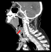
BACKGROUND Retropharyngeal hematoma is a relatively rare diagnosis that requires a high clinical suspicion and stabilization of the airway to prevent rapid deterioration. We report a case of a spontaneous retropharyngeal hematoma in an elderly patient with myelodysplastic syndrome and associated thrombocytopenia. CASE REPORT A 90-year-old man with myelodysplastic syndrome was brought to the Emergency Department with complaints of difficulty swallowing and muffled voice for 24 hours. Upon arrival, his vital signs and physical exam were unremarkable, except that when he was asked to take a sip of water, he could not swallow it. Complete blood count was remarkable for leukocytosis of 14.3×10³/mcL, hemoglobin of 9.0 gm/dL, and platelet count of 26×10³/mcL. Chest X-ray and lateral soft-tissue neck X-rays were grossly unremarkable. The patient was admitted for further evaluation and was scheduled for esophagogastroduodenoscopy. During intubation for esophagogastroduodenoscopy, the patient was noted to have significant airway narrowing. A subsequent CT scan revealed a 3×2×2 cm supraglottic hypodensity, thought to represent a retropharyngeal hematoma. The patient was transferred to the Intensive Care Unit (ICU) and received platelet transfusions. The ICU course was complicated by anemia, which necessitated transfusion of packed red blood cells. On hospital day 7, the patient reported resolution of his symptoms and was discharged home. CONCLUSIONS This case adds to the growing body of literature on spontaneous retropharyngeal hematomas. High clinical suspicion is warranted in patients who present with acute dysphagia, odynophagia, and dysphonia. Prompt imaging and airway management are vital in managing patients with this condition.
Conflict of interest statement
None.
Figures


Similar articles
-
Life-threatening airway obstruction due to retropharyngeal and cervicomediastinal hematomas following stellate ganglion block.Thorac Cardiovasc Surg. 2009 Aug;57(5):311-2. doi: 10.1055/s-2008-1038845. Epub 2009 Jul 23. Thorac Cardiovasc Surg. 2009. PMID: 19629898
-
[Retropharyngeal hematoma secondary to neck trauma--case report].Otolaryngol Pol. 2008;62(6):800-2. doi: 10.1016/S0030-6657(08)70366-4. Otolaryngol Pol. 2008. PMID: 19205538 Polish.
-
Anticoagulation and spontaneous retropharyngeal hematoma.J Emerg Med. 2003 May;24(4):389-94. doi: 10.1016/s0736-4679(03)00035-0. J Emerg Med. 2003. PMID: 12745040
-
Retropharyngeal hematoma secondary to minor blunt neck trauma: case report.Rev Bras Anestesiol. 2012 Sep-Oct;62(5):731-5. doi: 10.1016/S0034-7094(12)70171-X. Rev Bras Anestesiol. 2012. PMID: 22999405 Review.
-
Spontaneous retropharyngeal hemorrhage causing airway obstruction: a case report with a review of the literature.S D Med. 2006 Jul;59(7):295-7, 299. S D Med. 2006. PMID: 16895052 Review.
References
-
- Naqvi A, Hawwass D, Seto A. A tough pill to swallow-spontaneous retropharyngeal hematoma: a rare and unusual complication of rivaroxaban therapy. Cardiology and Angiology: An International Journal. 2015;4(4):156–59.
-
- Bloom DC, Haegen T, Keefe MA. Anticoagulation and spontaneous retropharyngeal hematoma. J Emerg Med. 2003;24(4):389–94. - PubMed
-
- Martí Gomar L, Gomar LM, Jiménez MG, Garcerán RM. Spontaneous retropharyngeal haematoma. Acta Otorrinolaringologica (English Edition) 2012;63(1):77–78. - PubMed
Publication types
MeSH terms
LinkOut - more resources
Full Text Sources
Other Literature Sources
Medical
Miscellaneous

