. 2018 Nov;11(6):1727-1742.
doi: 10.1038/s41385-018-0071-y. Epub 2018 Aug 16.

A novel role for C-C motif chemokine receptor 2 during infection with hypervirulent Mycobacterium tuberculosis

Affiliations

A novel role for C-C motif chemokine receptor 2 during infection with hypervirulent Mycobacterium tuberculosis

Micah D Dunlap et al. Mucosal Immunol. 2018 Nov.

Abstract

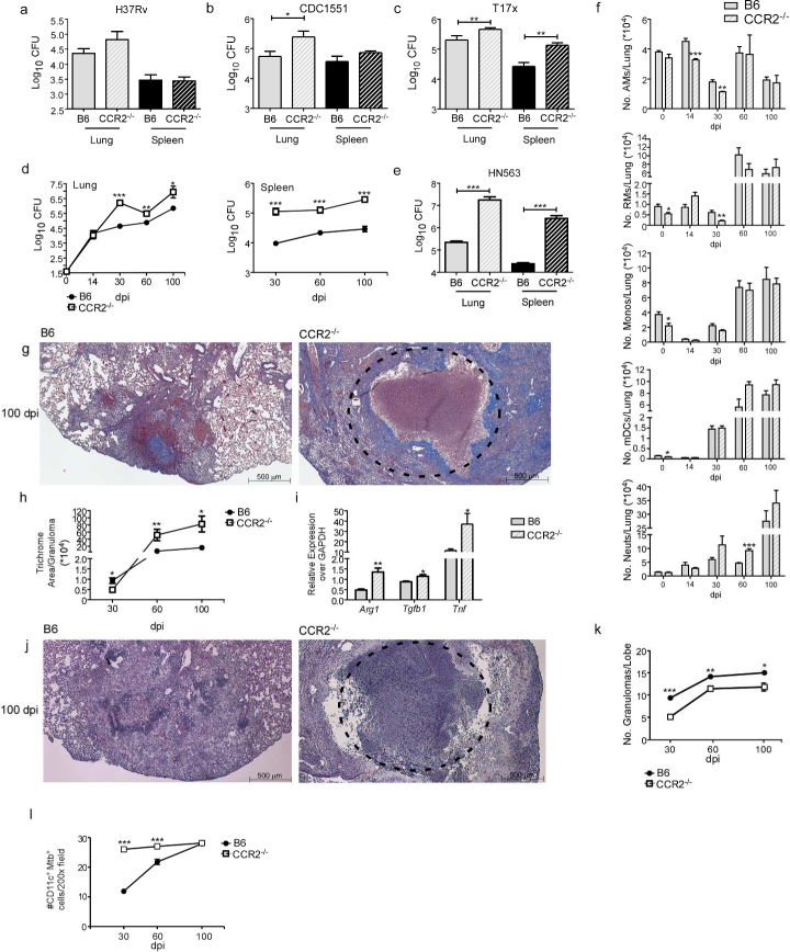
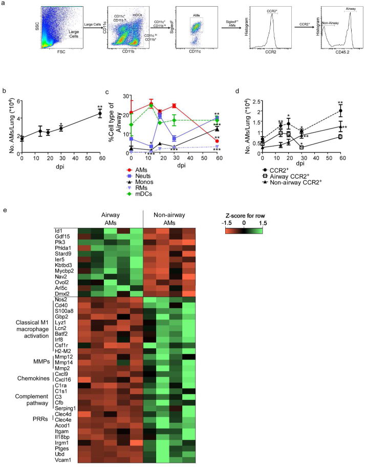
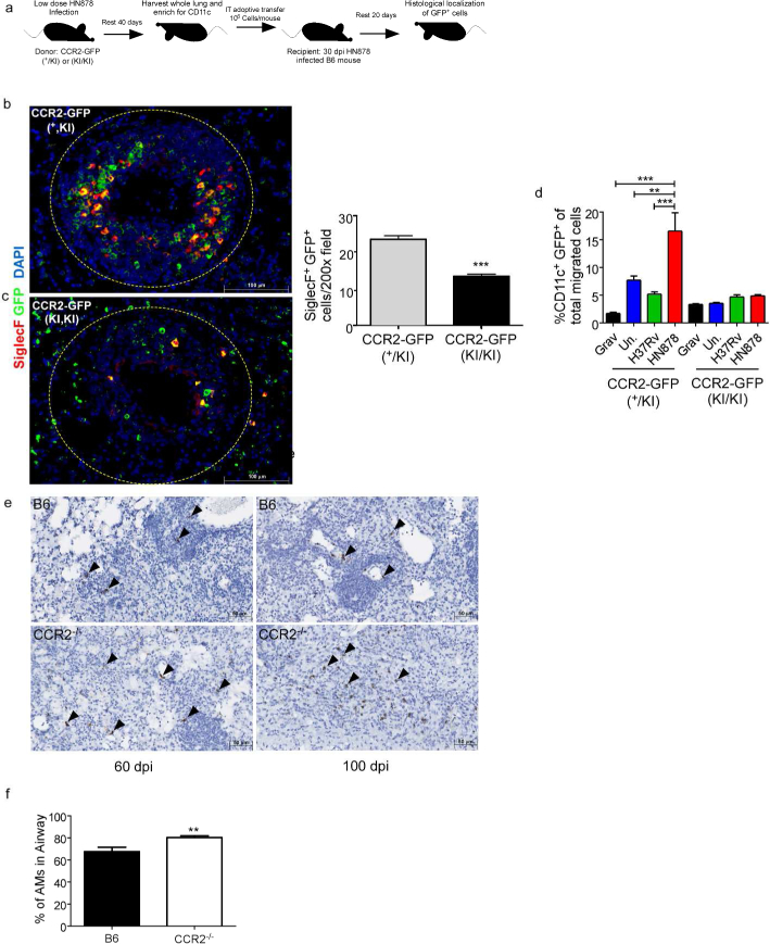
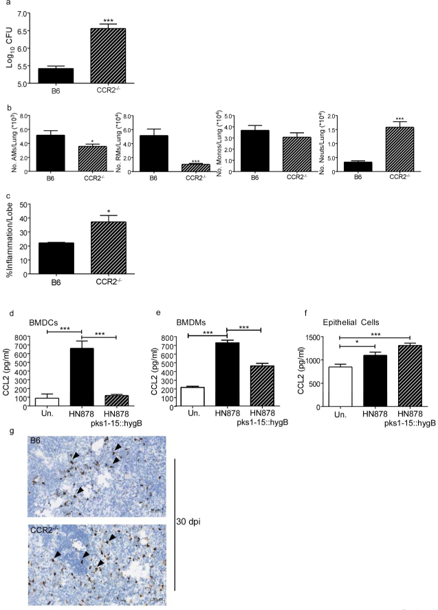
C-C motif chemokine receptor 2 (CCR2) is a major chemokine axis that recruits myeloid cells including monocytes and macrophages. Thus far, CCR2-/- mice have not been found to be susceptible to infection with Mycobacterium tuberculosis (Mtb). Here, using a prototype W-Beijing family lineage 2 Mtb strain, HN878, we show that CCR2-/- mice exhibit increased susceptibility to tuberculosis (TB). Following exposure to Mtb HN878, alveolar macrophages (AMs) are amongst the earliest cells infected. We show that AMs accumulate early in the airways following infection and express CCR2. During disease progression, CCR2-expressing AMs exit the airways and localize within the TB granulomas. RNA-sequencing of sorted airway and non-airway AMs from infected mice show distinct gene expression profiles, suggesting that upon exit from airways and localization within granulomas, AMs become classically activated. The absence of CCR2+ cells specifically at the time of AM egress from the airways resulted in enhanced susceptibility to Mtb infection. Furthermore, infection with an Mtb HN878 mutant lacking phenolic glycolipid (PGL) expression still resulted in increased susceptibility in CCR2-/- mice. Together, these data show a novel role for CCR2 in protective immunity against clinically relevant Mtb infections.


Conflict of interest statement

Conflict of Interest:

The authors declare no conflicts of interest.

The authors declare no competing financial interests.

Figures

Figure 1:
Figure 1:. CCR2−/− mice show increased susceptibility to low dose aerosol HN878 infection
B6 and CCR2−/− mice were aerosol-infected with ~100 CFU of (a) H37Rv, (b) CDC1551, (c) T17x, (d) HN878, or (e) HN563. Bacterial burden in the lung and spleen was determined by plating (a-c and e) at 30 dpi or (d) at different dpi. (f) Lung myeloid cell populations were enumerated in B6 and CCR2 −/− HN878-infected mice using flow cytometry at indicated dpi. (g-k) Pulmonary histology was assessed on FFPE lung sections from 30, 60 and 100 dpi samples stained with (g) Trichrome staining or (j) H&E staining. (h) Inflammatory area expressing collagen was quantified using Visiomorph image processing software to determine lung fibrosis.(i) RNA was extracted from B6 and CCR2−/− HN878-infected lungs and relative mRNA expression of specific genes was determined by qRT-PCR. Gapdh was used as internal control.(k) Inflammation was quantified using the morphometric tool of the Zeiss Axioplan microscope to determine the total number of granulomas per lobe. (l) The total number of CD11c+ Mtb containing cells per 200X was determined by counting in FFPE lung sections of B6 and CCR2−/− mice. AMs=Alveolar Macrophages, RMs=Recruited Macrophages, Monos=Monocytes, mDCs=Myeloid Dendritic Cells, Neuts=Neutrophils. n=5, (a-e) Student’s t-test between B6 and CCR2−/−, (f) 2-way ANOVA with Bonferroni’s post test. (h-l) Student’s t-test was used to determine differences per time point.
Figure 2:
Figure 2:. AMs are preferentially infected with HN878 and CCR2 expression on AMs is Mtb strain dependent
(a) The flow cytometry gating strategy for myeloid populations. Briefly, AMs were defined as CD11bCD11c+Siglec F+ cells. mDCs were defined as CD11b+CD11c+ cells. Neutrophils were defined as CD11b+CD11cGr-1hi cells, monocytes were defined as CD11b+CD11cGr-1lo cells, and recruited macrophages were defined as CD11b+CD11cGr-1 cells. Mtb-GFP+ cells were gated from individual subsets. (b) B6 mice were aerosol-infected with ~100 CFU of H37Rv-GFP or HN878-GFP and myeloid cell subsets infected with Mtb-GFP were determined by flow cytometry on 30 dpi (n=5). (c,d) B6 mice (n=5) were infected with aerosolized H37Rv or HN878 and the total number (c) and percentage (d) of CCR2+ myeloid lung cell populations were determined by flow cytometry and compared to uninfected controls. (e) Representative histograms of each subset displaying CCR2 expression by antibody staining compared to CCR2−/−. (f) Mean fluorescence intensity (MFI) of CCR2 expression on myeloid populations was normalized relative to MFI of unstained, uninfected controls. Un.=uninfected, AMs=Alveolar Macrophages, Monos=Monocytes, RMs=Recruited Macrophages, Neuts=Neutrophils, mDCs=Myeloid Dendritic Cells. (a-d) 2-Way ANOVA with Bonferroni posttest was used. (f) Student’s t-test.
Figure 3:
Figure 3:. Increased early CCR2 ligand expression is induced in the lung following HN878 infection
(a) RT-PCR analysis was performed at 21 and 30 dpi to determine mRNA expression for Ccl2,Ccl7, and Ccl12 in lungs of H37Rv- or HN878-infected mice (n=5 per group, per time point). (b)C10 epithelia, BMDCs, and BMDMs were cultured and infected with indicated Mtb strains at an MOI of 1 for 48 hours (n=6). Supernatants were analyzed by multiplex or ELISA assay forCCL2. (c-e) IKK2 fl/fl Sftpc-Cre mice and littermate controls were infected with HN878 for 14 (n=7) and 30 (n=5) dpi and the accumulation of (c) AMs, (d) neutrophils, and (e) RMs were calculated by flow cytometry. (f) Bacterial burden in the lung was determined by plating at 14 (n=7 per group) and 30 dpi (n=5 per group) in IKK2 fl/fl Sftpc-Cre mice and littermate controls. (g) Confocal microscopy of lung sections stained for E-cadherin (red) and CCL2 (green) in IKK2 fl/fl Sftpc-Cre mice and littermate controls. AMs=Alveolar Macrophages, Neuts=Neutrophils, RMs=Recruited Macrophages. (a) 2-way ANOVA with Bonferroni posttest (b) 1-way ANOVA with Tukey’s posttest. (c-f) Student’s t-test was used to compare between groups per time point.
Figure 4:
Figure 4:. CCR2 expression is required for AMs to egress from airways and localize within TB granulomas
Single cell lung suspensions from uninfected and infected mice (n=5) were prepared and (a) the gating strategy for airway and non-airway AMs is shown. The percentage and number of specific cell subsets with airway label CD45.2 delivered IT is shown. CD11c+CD11bloSiglecF+CD45.2+ cells were gated as airway AMs, while CD11c+CD11blo SiglecF+ CD45.2 cells were gated as non-airway AMs. (b) The total number of AMs over the time course of HN878 in B6 mice was determined by flow cytometry (n=5 per time point). (c) From total airway labelled cells (CD45.2+), the percentage of each myeloid cell type was determined in HN878-infected B6 mice by flow cytometry. (d) Total CCR2+ AMs, CCR2+ airway (CD45.2+) AMs, and CCR2+ non-airway (CD45.2) AMs over the course of HN878 infection in B6 mice were determined by flow cytometry. (e) Z-score Pearson correlation-based clustering of differentially expressed genes of interest (all 12 significantly differentially expressed genes in airway AMs, and 29 genes of functional interest that were higher in non-airway AMs). AMs=Alveolar Macrophages, Neuts=Neutrophils, Monos=Monocytes, RMs=Recruited Macrophages, mDCs=Myeloid Dendritic Cells, MMPs=matrix metallopeptidases, PRRs=pattern recognition receptors. (b-d) Each time point was compared to baseline d0 counts using Student’s t-test. (e) The gene expression levels of a subset of significantly differentially expressed genes between airway and non-airway AMs (according to DESeq).
Figure 5:
Figure 5:. CCR2 is required for AM localization within TB granulomas
(a) CD11c+ cells were purified from lungs of 30 dpi HN878-infected CCR2-GFP(+/KI) or CCR2-GFP(KI/KI) mice and 106 cells were IT transferred into B6 HN878-infected mice (n=5) at 30 dpi.(a-c) Lungs were harvested at 50 dpi and examined for localization of SiglecF+ GFP+ cells within TB granulomas using the morphometric tool of the Zeiss Axioplan microscope. (d) CCR2-GFP(+/KI) or CCR2-GFP(KI/KI) BMDMs were stimulated in vitro with 20 μg/mL irradiated Mtb HN878 for 24 hours. Migration towards uninfected, H37Rv- or HN878-infected epithelial cell supernatants was analyzed via transwell chemotaxis assays and flow cytometry (n=3). (e) Ccl2 mRNA localization was determined within FFPE lung sections from B6 and CCR2−/− HN878-infected using RNAScope in situ hybridization (ISH). Arrows point to Ccl2 mRNA localization(brown). (f) B6 and CCR2−/− mice (n=5) were infected with HN878 and percentage of AMs with airway label CD45.2 delivered IT was calculated on 14 dpi by flow cytometry. Grav=Gravity control, Un.=uninfected, AMs=Alveolar Macrophages. n=5 (b,c) Student’s t-test. n=3, (d) 2-Way ANOVA with Bonferroni posttest. (f) Student’s t-test.
Figure 6:
Figure 6:. Depletion of CCR2+ cells at the time of AM egress from airways increases susceptibility to HN878 infection
CCR2-DTR mice (n=4) were infected with HN878 and administered Dtx (a-c) IP at 12, 14, and 16 dpi or (d-f) at −1, 1, and 3 dpi. (a, d) Lung bacterial burden was determined by plating on 30 dpi. (b, e) Neutrophil, AM, monocyte and RM numbers were determined in PBS treated and Dtx treated CCR2-DTR mice at 30 dpi. (c, f) Pulmonary histology was assessed on FFPE lung sections stained with H&E, and inflammatory area was quantified using the morphometric tool of the Zeiss Axioplan microscope. (g) B6 (n=5) and CCR2−/− mice (n=8 per group) were infected with HN878, and CCR2−/− mice received either PBS or HN878-stimulated BMDMs delivered IT on 15 and 21 dpi. Lungs were harvested at 30 dpi and bacterial burden was determined by plating. Neuts=Neutrophils, AMs=Alveolar Macrophages, Monos=Monocytes, RMs=Recruited Macrophages. (a-f) Student’s t-test. (g) 1-way ANOVA with Tukey’s posttest.
Figure 7:
Figure 7:. Dependence on CCR2 for protective immunity to Mtb HN878 is not driven by PGL expression
B6 and CCR2−/− mice (n=5) were aerosol-infected with ~100 CFU HN878 pks1–15::hygB. (a) Lung bacterial burden was determined by plating on 30 dpi. (b) Lung myeloid cell populations of AMs, RMs, monocytes and neutrophils were enumerated in B6 and CCR2 −/− HN878 pks1–15:hygB infected mice using flow cytometry. (c) Pulmonary histology was assessed on FFPE lung sections stained with H&E, and inflammatory area was quantified using the morphometric tool of the Zeiss Axioplan microscope. (d-f) BMDCs, BMDMs, and C10 epithelial cells were cultured and infected with indicated Mtb strains at an MOI of 1 for 48 hours (n=5). Supernatants 34 were analyzed by multiplex or ELISA assay for CCL2. (g) Ccl2 mRNA localization was determined within FFPE lung sections from B6 and CCR2−/− HN878-infected using RNA Scope in situ hybridization (ISH). Arrows point to Ccl2 mRNA localization (brown). AMs=Alveolar Macrophages, RMs=Recruited Macrophages, Monos=Monocytes, Neuts=Neutrophils. (a-c) Student’s t-test. (d-f) 1-way ANOVA with Tukey’s posttest.

References

    1. Scott HM & Flynn JL Mycobacterium tuberculosis in chemokine receptor 2-deficient mice: influence of dose on disease progression. Infect. Immun 70, 5946–5954 (2002). - PMC - PubMed
    1. Samstein M et al. Essential yet limited role for CCR2(+) inflammatory monocytes during Mycobacterium tuberculosis-specific T cell priming. Elife 2, e01086, 10.7554/eLife.01086 (2013). - DOI - PMC - PubMed
    1. Domingo-Gonzalez R, Prince O, Cooper A & Khader SA Cytokines and Chemokines in Mycobacterium tuberculosis Infection. Microbiol Spectr 4, 10.1128/microbiolspec.TBTB2-0018-2016 (2016). - DOI - PMC - PubMed
    1. Tian G, Li X, Li H, Wang X & Cheng B Systematic meta-analysis of the association between monocyte chemoattractant protein-1 −2518A/G polymorphism and risk of tuberculosis. Genet. Mol. Res 14, 5501–5510, 10.4238/2015.May.25.1 (2015). - DOI - PubMed
    1. Reed MB et al. A glycolipid of hypervirulent tuberculosis strains that inhibits the innate immune response. Nature 431, 84–87, 10.1038/nature02837 (2004). - DOI - PubMed

Publication types

MeSH terms