. 2018 Aug 16;8(1):12272.
doi: 10.1038/s41598-018-30739-5.

Isolation and differential transcriptome of vascular smooth muscle cells and mid-capillary pericytes from the rat brain

Affiliations

Isolation and differential transcriptome of vascular smooth muscle cells and mid-capillary pericytes from the rat brain

Stéphanie Chasseigneaux et al. Sci Rep. .

Abstract

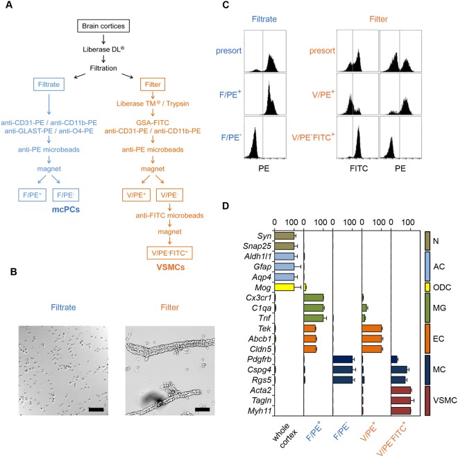
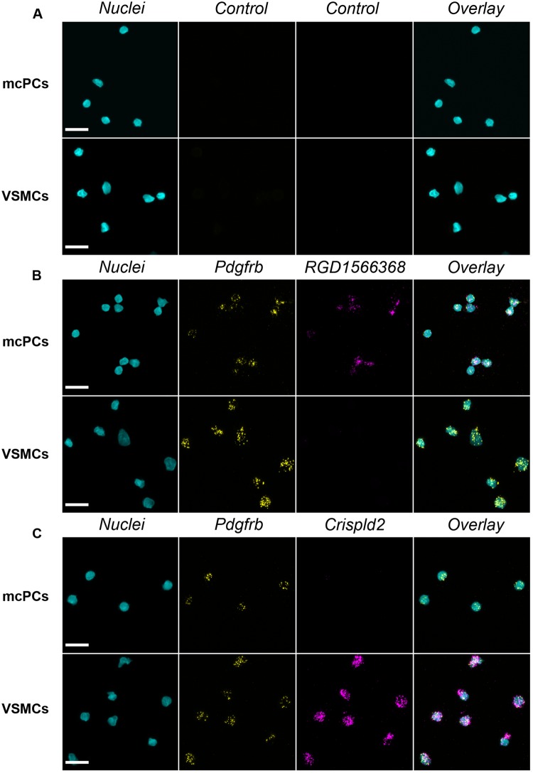
Brain mural cells form a heterogeneous family which significantly contributes to the maintenance of the blood-brain barrier and regulation of the cerebral blood flow. Current procedures to isolate them cannot specifically separate their distinct subtypes, in particular vascular smooth muscle cells (VSMCs) and mid-capillary pericytes (mcPCs), which differ among others by their expression of smooth muscle actin (SMA). We herein describe an innovative method allowing SMA+ VSMCs and SMA- mcPCs to be freshly isolated from the rat cerebral cortex. Using differential RNA-Seq analysis, we then reveal the specific gene expression profile of each subtype. Our results refine the current description of the role of VSMCs in parenchymal cortical arterioles at the molecular level and provide a unique platform to identify the molecular mechanisms underlying the specific functions of mcPCs in the brain vasculature.


Conflict of interest statement

The authors declare no competing interests.

Figures

Figure 1
Figure 1
VSMCs and mcPCs can be selectively sorted from the rat cerebral cortex. (A) Simplified flowchart of the procedure used to isolate brain mural cells from the mid-capillary bed (Filtrate) and parenchymal arterioles (Filter). mcPCs and VSMCs are recovered in the fractions F/PE and V/PEFITC+, respectively. (B) Phase contrast microscopy of the filtrate and arterioles retained on the 10 µm mesh filter (scale bar 50 µm). (C) Flow cytometry analysis of magnetically sorted cells. PE fluorescence distribution of magnetically labeled cells from the filtrate before (presort) and after (F/PE+, F/PE) magnetic sorting (left panel). FITC and PE fluorescence distributions of magnetically labeled cells from dissociated arterioles before (presort) and after (V/PE+, V/PE-FITC+) magnetic sorting (right panel). (D) qRT-PCR analysis confirms the nature and purity of magnetically sorted mural cells. Whole cortex and sorted fractions were analyzed for the relative expression of the indicated specific markers of neurons (N), astrocytes (AC), oligodendrocytes (ODC), microglia (MG), endothelial cells (EC), mural cells (MC) and vascular smooth muscle cells (VSMC). For each gene, values are normalized to the highest value across all samples (mean ± SEM from three independent experiments).
Figure 2
Figure 2
Isolated VSMCs and mcPCs display distinct RNA-Seq transcriptomic signatures. (A,B) These heatmaps include the 12,203 genes of the final dataset and compare the samples (mcPC1-3 and VSMC1-3) from three independent experiments. (A) The heatmap of the sample-to-sample euclidian distances showing high correlation among biological replicates but low correlation between mcPCs and VSMCs. (B) The heatmap of standard deviations from mean normalized count values showing that mcPCs and VSMCs display distinct gene expression profiles. (C) Differential expression for usual mural markers (LFC is positive for genes enriched in mcPCs, negative for genes enriched in VSMCs). (D) Gene Ontology pathways enriched among the genes highly overexpressed in VSMCs (LFC < −2) or mcPCs (LFC > 2). Only the 20 most significantly enriched pathways are shown.
Figure 3
Figure 3
FISH confirms the differential expression of RGD1566368 and Crispld2 transcripts in freshly isolated mcPCs and VSMCs. (A) A control probe gives virtually no signal in both cell types. (B) Pdgfrb (yellow)/RGD1566368 (magenta) double FISH. mcPCs and VSMCs are positive for Pdgfrb but only mcPCs are positive for RGD1566368. (C) Pdgfrb (yellow)/Crispld2 (magenta) double FISH. mcPCs and VSMCs are positive for Pdgfrb but only VSMCs are positive for Crispld2. (AC) Hoechst staining (nuclei) in cyan, scale bar 10 µm.
Figure 4
Figure 4
FISH confirms the differential expression of RGD1566368 and Crispld2 transcripts in mural cells of mechanically isolated brain vessels. (A) A control probe gives virtually no signal. (B) Pdgfrb (yellow)/RGD1566368 (magenta) double FISH. Both mcPCs in capillaries (arrowheads) and VSMCs in arterioles (arrow) are positive for Pdgfrb but only mcPCs are positive for RGD1566368. (C) Pdgfrb (yellow)/Crispld2 (magenta) double FISH. Both mcPCs in capillaries (arrowheads) and VSMCs in arterioles (arrow) are positive for Pdgfrb but only VSMCs are positive for Crispld2. A,B,C, DIC image in gray, Hoechst staining (nuclei) in cyan, scale bar 20 µm.

References

    1. Armulik A, Genové G, Betsholtz C. Pericytes: developmental, physiological, and pathological perspectives, problems, and promises. Dev. Cell. 2011;21:193–215. doi: 10.1016/j.devcel.2011.07.001. - DOI - PubMed
    1. Etchevers HC, Vincent C, Le Douarin NM, Couly GF. The cephalic neural crest provides pericytes and smooth muscle cells to all blood vessels of the face and forebrain. Dev. Camb. Engl. 2001;128:1059–1068. - PubMed
    1. Korn J, Christ B, Kurz H. Neuroectodermal origin of brain pericytes and vascular smooth muscle cells. J. Comp. Neurol. 2002;442:78–88. doi: 10.1002/cne.1423. - DOI - PubMed
    1. Grant, R. I. et al. Organizational hierarchy and structural diversity of microvascular pericytes in adult mouse cortex. J. Cereb. Blood Flow Metab. Off. J. Int. Soc. Cereb. Blood Flow Metab. 271678X17732229, 10.1177/0271678X17732229 (2017). - PMC - PubMed
    1. Hartmann DA, et al. Pericyte structure and distribution in the cerebral cortex revealed by high-resolution imaging of transgenic mice. Neurophotonics. 2015;2:041402. doi: 10.1117/1.NPh.2.4.041402. - DOI - PMC - PubMed

Publication types