Review
. 2018 Sep;17(9):660-688.
doi: 10.1038/nrd.2018.109. Epub 2018 Aug 17.

Promoting the clearance of neurotoxic proteins in neurodegenerative disorders of ageing

Affiliations
Review

Promoting the clearance of neurotoxic proteins in neurodegenerative disorders of ageing

Barry Boland et al. Nat Rev Drug Discov. 2018 Sep.

Abstract

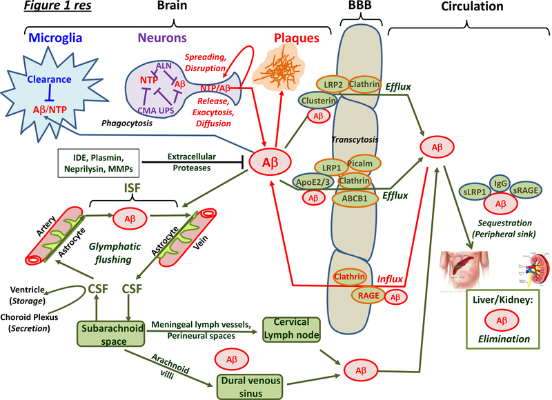
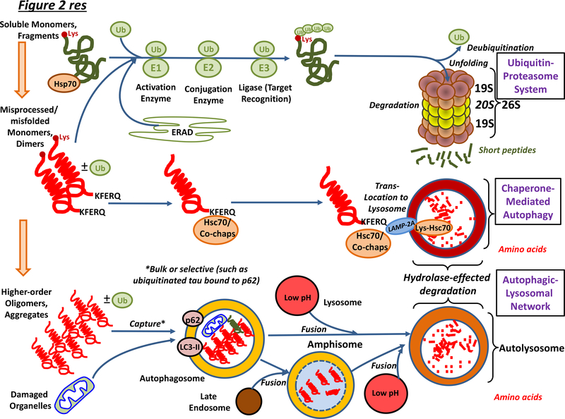
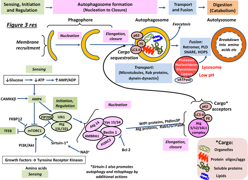
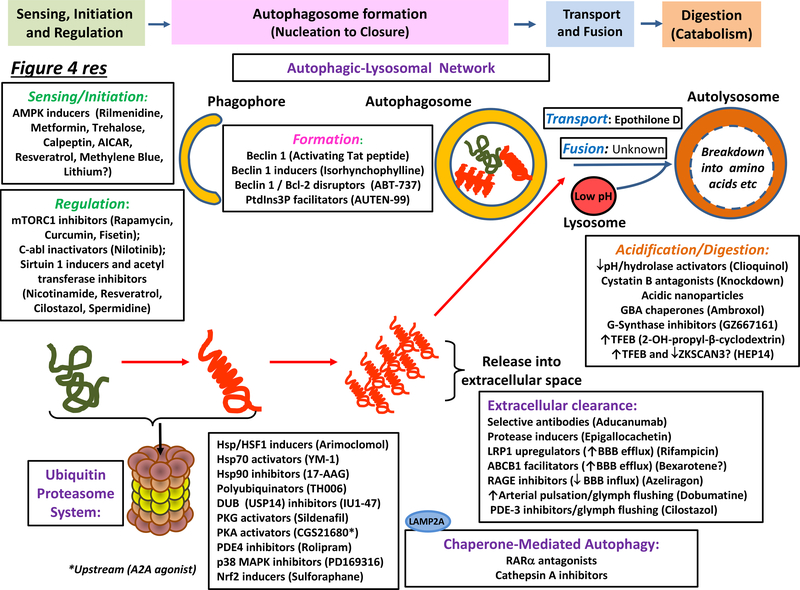
Neurodegenerative disorders of ageing (NDAs) such as Alzheimer disease, Parkinson disease, frontotemporal dementia, Huntington disease and amyotrophic lateral sclerosis represent a major socio-economic challenge in view of their high prevalence yet poor treatment. They are often called 'proteinopathies' owing to the presence of misfolded and aggregated proteins that lose their physiological roles and acquire neurotoxic properties. One reason underlying the accumulation and spread of oligomeric forms of neurotoxic proteins is insufficient clearance by the autophagic-lysosomal network. Several other clearance pathways are also compromised in NDAs: chaperone-mediated autophagy, the ubiquitin-proteasome system, extracellular clearance by proteases and extrusion into the circulation via the blood-brain barrier and glymphatic system. This article focuses on emerging mechanisms for promoting the clearance of neurotoxic proteins, a strategy that may curtail the onset and slow the progression of NDAs.


Figures

Figure 1 |
Figure 1 |. Overview of intracelluar and extracellular mechanisms for the clearance of neurotoxic proteins from the brain.
Neurotoxic proteins (NTPs) are eliminated by a broad suite of specific and non-specific mechanisms in neurons, glial cells and endothelial/vascular smooth muscle cells of vessels. The three major modes of intracellular clearance — the autophagic–lysosomal network (ALN), chaperone-mediated autophagy (CMA) and the ubiquitin–proteasome system (UPS) — are shown for neurons but they are also active in other cells such as microglia. Under conditions of inflammation, proteasomal β-subunits in glia are switched and substrate specificity changes: the precise role of these ‘immunoproteasomes’ — specialized in peptide production for antigen presentation — for neurotoxic protein elimination in NDAs is debated. Clearance also occurs in the extracellular space, the interstitial fluid (ISF) of the brain parenchyma that surrounds neurons, and the cerebrospinal fluid (CSF) with which the ISF exchanges. Intraneuronal mechanisms of clearance are illustrated for NTPs in general, but only Aβ42 is shown for extracellular clearance, since the vast majority of currently available data is for this NTP. Extracellular pools of NTPs are derived from passive diffusion, active release from terminals, extrusion by exocytosis, and dispersion upon cell death. NTPs disrupt neuronal and synaptic function and are taken up by other neurons and glial cells (‘spreading’). Therapeutically relevant proteases degrading NTPs include endothelin-converting enzyme and insulin degrading enzyme (IDE) (mainly cytosolic), neprilysin and matrix metalloproteinases (MMP) (intracellular and extracellular), and plasmin (mainly extracellular). NTPs that escape glial capture and proteases are driven into the circulation. First, blood–brain barrier (BBB) localised receptors and transporters actively eject them into the blood, including P-glycoproteins such as ABCB1 transporters and low-density lipoprotein receptor related protein 1 (LRP1). Conversely, the receptor for advanced glycation end-product (RAGE) receptor returns Aβ into the CNS. Similar mechanisms operate at the blood–CSF barrier in the choroid plexus; for example, LRP2 transfer of transthyretin-bound Aβ from CSF into blood. Second, transfer of NTPs to the periphery is mediated through the glymphatic system. CSF runs along the peri-arterial space, transverses aquaporin 4 receptor-bearing circumvascular astrocytes to enter the ISF. Convective flow driven by arterial pulsing flushes NTPs via glial cells and the peri-venous space back into the CSF. Glymphatic-cleared, CSF-derived NTPs mainly reach the circulation mainly via the cervical lymph nodes, but also via the dural venous sinus. Within the blood, specific proteins sequester Aβ, such as the soluble fragment of LRP1 and immunoglobulins (IgG). NTPs are ultimately eliminated in the kidneys and liver. Abbreviation not in main text or above: s, soluble.
Figure 2 |
Figure 2 |. Overview of intracellular mechanisms for the elimination of neurotoxic proteins from neurons and other classes of cell in the brain.
Within neurons and other classes of cell, the UPS and CMA clear non-aggregated forms of neurotoxic protein, and the UPS also deals with substrates of endoplasmic reticulum-associated degradation (ERAD) of incorrectly-folded proteins. Proteins destined for the proteasome are poly-ubiquinated and guided to the proteasome by chaperones. They are deubiquinated by Rpn11 once committed to entering the proteosome pore: other deubiquitinases such as USP14 may rescue them before entry. Unfolding is followed by degradation. The CMA operates on proteins bearing a KFERQ-like motif. This sequence is found in, for example, tau but not Aβ. Hsc70 recognises the KFERQ sequence and, together with co-chaperones, transports the protein to the LAMP2A receptor on lysosomes: LAMP2A then coordinates protein translocation into the lumen. The ALN is the major system for removing misfolded, higher-order, aggregated proteins as well as damaged organelles. Autophagosomes bearing cargo fuse with acidic lysosomes, leading to degradation of contents. In addition, some autophagosomes fuse with late endosomes. The resultant amphisomes then likewise fuse with lysosomes. See also Figure 3. Abbreviation not in main text: Co-chap, co-chaperone; Lys, lysine and Ub, ubiquitin,
Figure 3 |
Figure 3 |. Organization, operation and regulation of the autophagic–lysosomal network.
The top part of the figure illustrates the sequence of steps associated with operation of the ALN, while the bottom part shows the main regulatory proteins involved, focusing on potential targets for pharmacotherapy. ‘Sensing’ — both extrinsic (for example, glucose levels) and intrinsic (e.g. for example, ATP/AMP levels) — can determine whether or not autophagy is initiated by activation of AMPK and/or inhibition of mTORC1, which leads to TFEB-driven transcription of ALN-requisite proteins. The pre-autophagosome (phagophore) structure first emerges from diverse membrane sources, and its formation is promoted by Atg9 (not shown). Nucleation is accomplished with the help of a complex cluster of proteins. Phosphatidylinositol-3-phosphate (PtdIns3P) is recognised by WIPI (WD-repeat-protein-interacting-with-phosphoInositides) proteins that help induce autophagosome elongation in association with several classes of Atg protein and small GTPases such as Rab5. With the aid of LC3 and cargo acceptors, autophagosomes take up cytoplasmic material such as aggregated proteins and dysfunctional mitochondria (Box 2). Autophagosomes and other autophagic vesicles are transported with the help of dynactin and dynein along microtubules towards acidic lysosomes. Autophagosomes fuse with lysosomes containing resident hydrolases that degrade their contents into amino acids, sugars and lipids for recycling. Exosomal release/secretion of neurotoxic proteins (“exocytosis)”) may occur upon reduced ALN flux and accumulation of autophagosomes. For details, see main text. Abbreviations not in main text or Glossary: FIP, family interacting protein; HOPS; Homotypic fusion and protein sorting complex; NAD+, nicotinamide adenine dinucleotide; PE, phosphoethanolamine; PI3K/Akt: phosphoinositol-3-kinase/atypical kinase and PLD, phospholipase D.
Figure 4 |
Figure 4 |. Major molecular sites of action of agents that enhance neurotoxic protein clearance in neurodegenerative disorders of aging.
Representative agents are shown for diverse modes of intracellular (the autophagic–lysosomal network (ALN) and the ubiquitin–proteasome system (UPS), extracellular (immunotherapy and protease-driven) and vascular (blood–brain barrier (BBB) extrusion and glymphatic) clearance. The principal loci of drug actions are depicted, yet precise mechanisms of action remain to be more fully deciphered for many drugs while several agents like resveratrol act at multiple sites (main text). As illustrated, a broad range of drugs exert their actions via AMPK, mTORC1 or sirtuin 1 (which also influences downstream events such as autophagosome formation). Some agents exert their effects via other components of the ALN, up to and including lysosomal catabolism. In addition, ambroxol acts as a chaperone to help transport β−glucocerebrosidase to lysosomes. Diverse class of agent likewise promote UPS activity, including chaperones that assist in protein refolding and triage, modulators of proteasomal phosphorylation, and agents acting via the transcription factor, Nrf2, to induce coordinated synthesis of proteasomal subunits. Extraneuronal clearance of full-length, truncated, post-translationally-modified, monomeric and/or higher-order neurotoxic proteins can be promoted by: stimulating proteases like neprilysin; immunotherapies targeting specific neurotoxic proteins; and increasing BBB-mediated and glymphatic extrusion into the circulation. For details, see main text. Abbreviations not in main text or Figure 3: AT, acetyl transferase; DUB, deubiquitinase; GBA; β−glucocerebrosidase; G-synthase, glucoceramide synthase; PDE, phosphodiesterase; PKA/G, protein kinases A/G and RAR, retinoid acid receptor.

References

    1. Menzies FM et al. Autophagy and Neurodegeneration: Pathogenic Mechanisms and Therapeutic Opportunities. Neuron 93, 1015–1034, doi:10.1016/j.neuron.2017.01.022 (2017). - DOI - PubMed
    1. Ciechanover A & Kwon YT Protein Quality Control by Molecular Chaperones in Neurodegeneration. Front Neurosci 11, 185, doi:10.3389/fnins.2017.00185 (2017). - DOI - PMC - PubMed
    1. Dikic I Proteasomal and Autophagic Degradation Systems. Annu Rev Biochem 86, 193–224, doi:10.1146/annurev-biochem-061516-044908 (2017). - DOI - PubMed
    1. Galluzzi L et al. Molecular definitions of autophagy and related processes. EMBO J 36, 1811–1836, doi:10.15252/embj.201796697 (2017). - DOI - PMC - PubMed
    1. Galluzzi L, Bravo-San Pedro JM, Levine B, Green DR & Kroemer G Pharmacological modulation of autophagy: therapeutic potential and persisting obstacles. Nat Rev Drug Discov, 16, 487–511. doi:10.1038/nrd.2017.22 (2017). - DOI - PMC - PubMed

Publication types