

Early Imaging Biomarker of Myocardial Glucose Adaptations in High-Fat-Diet-Induced Insulin Resistance Model by Using 18F-FDG PET and [U-13C]glucose Nuclear Magnetic Resonance Tracer
- PMID: 30116165
- PMCID: PMC6079607
- DOI: 10.1155/2018/8751267
Early Imaging Biomarker of Myocardial Glucose Adaptations in High-Fat-Diet-Induced Insulin Resistance Model by Using 18F-FDG PET and [U-13C]glucose Nuclear Magnetic Resonance Tracer
Abstract
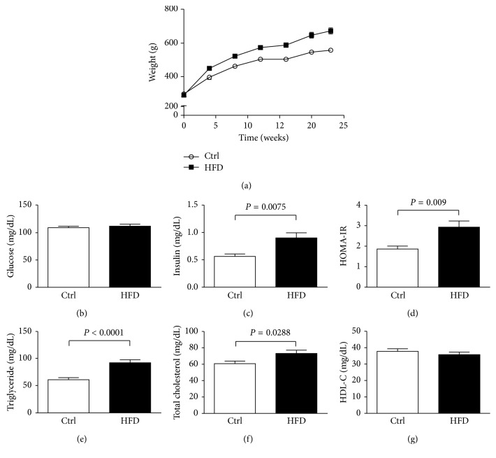
Background: High-fat diet (HFD) induces systemic insulin resistance leading to myocardial dysfunction. We aim to characterize the early adaptations of myocardial glucose utility to HFD-induced insulin resistance.
Methods: Male Sprague-Dawley rats were assigned into two groups, fed a regular chow diet or HFD ad libitum for 10 weeks. We used in vivo imaging of cardiac magnetic resonance (CMR), 18F-FDG PET, and ex vivo nuclear magnetic resonance (NMR) metabolomic analysis for the carbon-13-labeled glucose ([U-13C]Glc) perfused myocardium.
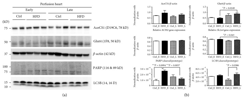
Results: As compared with controls, HFD rats had a higher ejection fraction and a smaller left ventricular end-systolic volume (P < 0.05), with SUVmax of myocardium on 18F-FDG PET significantly increased in 4 weeks (P < 0.005). The [U-13C]Glc probed the increased glucose uptake being metabolized into pyruvate and acetyl-CoA, undergoing oxidative phosphorylation via the tricarboxylic acid (TCA) cycle, and then synthesized into glutamic acid and glutamine, associated with overexpressed LC3B (P < 0.05).
Conclusions: HFD-induced IR associated with increased glucose utility undergoing oxidative phosphorylation via the TCA cycle in the myocardium is supported by overexpression of glucose transporter, acetyl-CoA synthase. Noninvasive imaging biomarker has potentials in detecting the metabolic perturbations prior to the decline of the left ventricular function.
Figures


Similar articles
-
Glucose Metabolism as a Pre-clinical Biomarker for the Golden Retriever Model of Duchenne Muscular Dystrophy.Mol Imaging Biol. 2018 Oct;20(5):780-788. doi: 10.1007/s11307-018-1174-2. Mol Imaging Biol. 2018. PMID: 29508262 Free PMC article.
-
In vivo cardiac glucose metabolism in the high-fat fed mouse: Comparison of euglycemic-hyperinsulinemic clamp derived measures of glucose uptake with a dynamic metabolomic flux profiling approach.Biochem Biophys Res Commun. 2015 Aug 7;463(4):818-24. doi: 10.1016/j.bbrc.2015.06.019. Epub 2015 Jun 15. Biochem Biophys Res Commun. 2015. PMID: 26086096
-
Role of Mitochondrial Oxidative Stress in Glucose Tolerance, Insulin Resistance, and Cardiac Diastolic Dysfunction.J Am Heart Assoc. 2016 May 5;5(5):e003046. doi: 10.1161/JAHA.115.003046. J Am Heart Assoc. 2016. PMID: 27151515 Free PMC article.
-
Imaging of cardiac metabolism using radiolabelled glucose, fatty acids and acetate.Coron Artery Dis. 2001 Feb;12 Suppl 1:S12-8. Coron Artery Dis. 2001. PMID: 11286301 Review.
-
Strategies for Imaging Metabolic Remodeling of the Heart in Obesity and Heart Failure.Curr Cardiol Rep. 2022 Apr;24(4):327-335. doi: 10.1007/s11886-022-01650-3. Epub 2022 Feb 2. Curr Cardiol Rep. 2022. PMID: 35107704 Free PMC article. Review.
Cited by
-
The role of (18F)-fluoro-D-glucose positron emission tomography/computed tomography in the surveillance of abnormal myocardial energy metabolism and cardiac dysfunction in a rat model of cardiopulmonary resuscitation.Diagn Interv Radiol. 2023 May 31;29(3):548-554. doi: 10.4274/dir.2023.221932. Epub 2023 May 8. Diagn Interv Radiol. 2023. PMID: 37154799 Free PMC article.
-
Changes in Cardiac Metabolism in Prediabetes.Biomolecules. 2021 Nov 12;11(11):1680. doi: 10.3390/biom11111680. Biomolecules. 2021. PMID: 34827678 Free PMC article. Review.
-
Regulation of energy metabolism by combination therapy attenuates cardiac metabolic remodeling in heart failure.Int J Biol Sci. 2020 Oct 16;16(16):3133-3148. doi: 10.7150/ijbs.49520. eCollection 2020. Int J Biol Sci. 2020. PMID: 33162820 Free PMC article.
References
Publication types
MeSH terms
Substances
LinkOut - more resources
Full Text Sources
Other Literature Sources
Research Materials
