

Entry points of nutrient arteries at risk during osteotomy of the lesser metatarsals: a fresh cadaveric study
- PMID: 30116305
- PMCID: PMC6083557
- DOI: 10.1186/s13047-018-0288-8
Entry points of nutrient arteries at risk during osteotomy of the lesser metatarsals: a fresh cadaveric study
Abstract
Background: Osteotomies of the lesser (second to fourth) metatarsals are often used to correct forefoot deformities. However, certain areas of the lesser metatarsals where arteries may be prone to damage during surgery, and the resulting nonunion and delayed union could cause serious problems. This study sought to identify the nutrient arteries of the lesser metatarsals and to determine how osteotomy could injure these vessels.
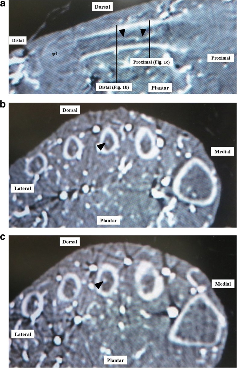
Methods: Enhanced computed tomography scans of 21 ft (male, n = 10; female, n = 11; mean age 78.6 years at the time of death) were assessed. Twenty-one lower limbs in 21 cadaveric specimens were injected with barium via the external iliac artery, and the points at which the nutrient arteries entered the lesser metatarsals were identified on axial and coronal images.
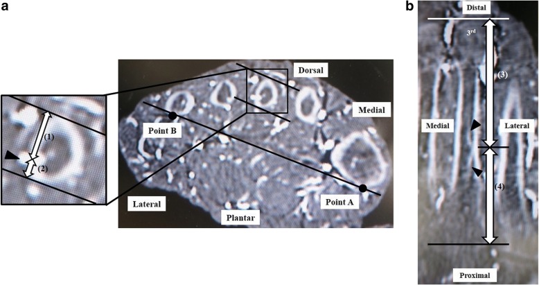
Results: Each nutrient artery entered the lateral or medial plantar aspect of the lesser metatarsal in the middle third (just proximal to the middle point of the metatarsal) or proximal third obliquely from a distal direction. The mean ± standard deviation (SD) distances from the dorsal plane of the second, third, and fourth metatarsals to the point of entry of the nutrient artery in the axial plane were 8.2 ± 1.5, 7.6 ± 1.2, and 7.6 ± 1.5 mm, respectively. The mean ± SD distances from the distal epiphysis to the point of entry of the nutrient artery into the second, third, and fourth metatarsals in the coronal plane were 3.3 ± 1.1, 3.1 ± 1.0, and 2.8 ± 1.2 mm, respectively. The mean ± SD distances from the distal epiphysis to the point of entry of the nutrient artery into the second, third, and fourth metatarsals in the coronal plane were 46.0 ± 5.2, 40.9 ± 2.6, and 39.1 ± 3.7 mm, respectively. The mean ± SD distances from the proximal epiphysis to the entry point of the nutrient artery into the second, third, and fourth metatarsals in the coronal plane were 23.8 ± 4.7, 25.8 ± 4.3, and 25.0 ± 3.2 mm, respectively.
Conclusions: Distal metatarsal osteotomies might be safer than shaft or proximal osteotomy to avoid disruption of the nutrient artery, leading to delayed consolidation of the osteotomy and nonunion.
Keywords: Cadaver; Lesser metatarsal; Nutrient artery; Osteotomy.
Conflict of interest statement
This study was approved by the research board of Tokushima University Hospital (reference no. 2258). Not applicable. The authors declare that they have no competing interests. Springer Nature remains neutral with regard to jurisdictional claims in published maps and institutional affiliations.
Figures


References
-
- Spence KF, O’Connell SJ, Kenzora JE. Proximal metatarsal segmental resection: a treatment for intractable plantar keratosis. Orthopedics. 1990;13:741–747. - PubMed
MeSH terms
LinkOut - more resources
Full Text Sources
Other Literature Sources
