

Evaluation of Threshold Values for Root Canal Filling Voids in Micro-CT and Nano-CT Images
- PMID: 30116470
- PMCID: PMC6079325
- DOI: 10.1155/2018/9437569
Evaluation of Threshold Values for Root Canal Filling Voids in Micro-CT and Nano-CT Images
Abstract
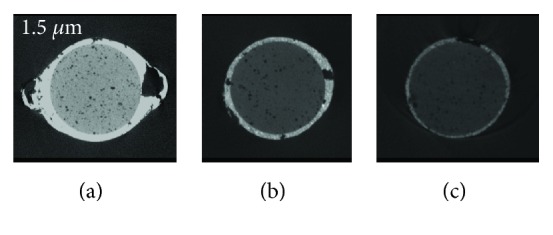
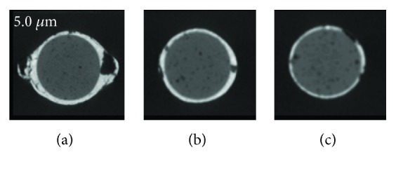
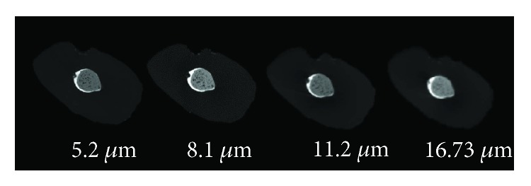
While several materials and techniques have been used to assess the quality of root canal fillings in micro-CT images, the lack of standardization in scanning protocols has produced conflicting results. Hence, the aim of this study was to determine a cutoff voxel size value for the assessment of root canal filling voids in micro-CT and nano-CT images. Twenty freshly extracted mandibular central incisors were used. Root canals were prepared with nickel titanium files to an ISO size 40/0.06 taper and then filled with a single cone (40/0.06 taper) and AH Plus sealer. The teeth were scanned with different voxel sizes with either micro-CT (5.2, 8.1, 11.2, and 16.73 μm) or nano-CT (1.5 and 5.0 μm) equipment. Images were reconstructed and analyzed with the NRecon and CTAn software. Void proportion and void volume were calculated for each tooth in the apical, middle, and coronal thirds of the root canal. Kruskal-Wallis and post hoc Mann-Whitney U tests were performed with a significance level of 5%. In micro-CT images, significantly different results were detected among the tested voxel sizes for void proportion and void volume, whereas no such differences were found in nano-CT images (p > 0.05). Micro-CT images showed higher void numbers over the entire root length, with statistically significant differences between the voxel size of 16.73 μm and the other sizes (p < 0.05). The values of the different nano-CT voxel sizes did not significantly differ from those of the micro-CT (5.2, 8.1, and 11.2 μm), except for the voxel size of 16.73 μm (p < 0.05). All tested voxel sizes enabled the detection of root canal filling voids except for the voxel size of 16.73 μm. Bearing in mind the limitations of this study, it seems that a voxel size of 11.2 μm can be used as a reliable cutoff value for the assessment of root canal filling voids in micro-CT imaging.
Figures
Similar articles
-
3D micro-CT analysis of the interface voids associated with Thermafil root fillings used with AH Plus or a flowable MTA sealer.Int Endod J. 2013 Mar;46(3):253-63. doi: 10.1111/j.1365-2591.2012.02124.x. Epub 2012 Oct 8. Int Endod J. 2013. PMID: 23039158 Clinical Trial.
-
Evaluation of root canal sealer filling quality using a single-cone technique in oval shaped canals: An In vitro Micro-CT study.Scanning. 2016 Mar-Apr;38(2):133-40. doi: 10.1002/sca.21249. Epub 2015 Jul 30. Scanning. 2016. PMID: 26228657
-
Micro-CT and nano-CT analysis of filling quality of three different endodontic sealers.Dentomaxillofac Radiol. 2017 Dec;46(8):20170223. doi: 10.1259/dmfr.20170223. Epub 2017 Nov 16. Dentomaxillofac Radiol. 2017. PMID: 28845679 Free PMC article.
-
3-dimensional Analysis and Literature Review of the Root Canal Morphology and Physiological Foramen Geometry of 125 Mandibular Incisors by Means of Micro-Computed Tomography in a German Population.J Endod. 2020 Feb;46(2):184-191. doi: 10.1016/j.joen.2019.11.006. Epub 2019 Dec 27. J Endod. 2020. PMID: 31889585 Review.
-
State of the art of Micro-CT applications in dental research.Int J Oral Sci. 2009 Dec;1(4):177-88. doi: 10.4248/IJOS09031. Int J Oral Sci. 2009. PMID: 20690421 Free PMC article. Review.
Cited by
-
Evaluation Algorithm of Root Canal Shape Based on Steklov Spectrum Analysis.Comput Math Methods Med. 2019 Nov 3;2019:4830914. doi: 10.1155/2019/4830914. eCollection 2019. Comput Math Methods Med. 2019. PMID: 31885681 Free PMC article.
-
How image-processing parameters can influence the assessment of dental materials using micro-CT.Imaging Sci Dent. 2020 Jun;50(2):161-168. doi: 10.5624/isd.2020.50.2.161. Epub 2020 Jun 18. Imaging Sci Dent. 2020. PMID: 32601591 Free PMC article.
-
Estimation of root canal conicity of deciduous canines evaluated by nano-CT.Eur Arch Paediatr Dent. 2023 Jun;24(3):335-342. doi: 10.1007/s40368-023-00809-y. Epub 2023 Jun 12. Eur Arch Paediatr Dent. 2023. PMID: 37306868
-
The Value of Micro-CT in the Diagnosis of Lung Carcinoma: A Radio-Histopathological Perspective.Diagnostics (Basel). 2023 Oct 20;13(20):3262. doi: 10.3390/diagnostics13203262. Diagnostics (Basel). 2023. PMID: 37892083 Free PMC article. Review.
-
Laboratory simulation of longitudinally cracked teeth using the step-stress cyclic loading method.Int Endod J. 2021 Sep;54(9):1638-1646. doi: 10.1111/iej.13530. Epub 2021 May 5. Int Endod J. 2021. PMID: 33852743 Free PMC article. Clinical Trial.
References
-
- Eskandarinezhad M., Shahveghar-Asl N., Sharghi R., et al. Sealing efficacy of mineral trioxide aggregate with and without nanosilver for root end filling: an in-vitro bacterial leakage study. Journal of Clinical and Experimental Dentistry. 2017;9(1):e27–33. doi: 10.4317/jced.53070. - DOI - PMC - PubMed
MeSH terms
LinkOut - more resources
Full Text Sources
Other Literature Sources
