

Role of the Scaffold Protein MIM in the Actin-Dependent Regulation of Epithelial Sodium Channels (ENaC)
- PMID: 30116621
- PMCID: PMC6087825
Role of the Scaffold Protein MIM in the Actin-Dependent Regulation of Epithelial Sodium Channels (ENaC)
Abstract
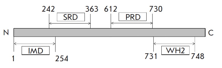
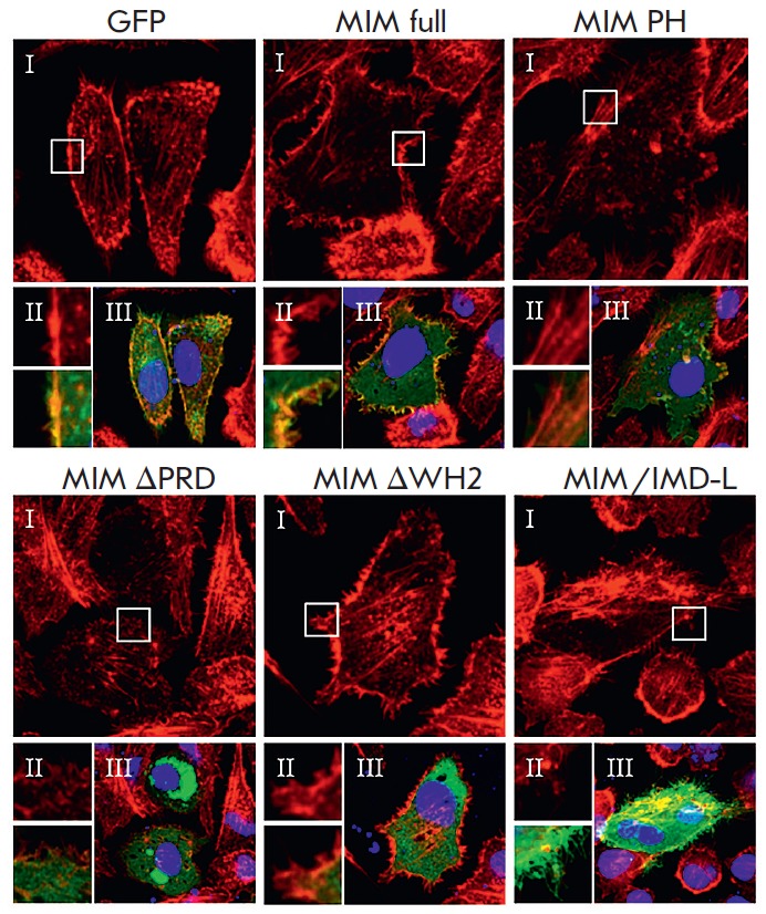
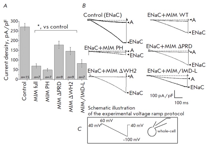
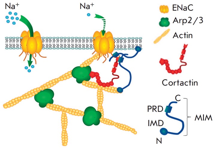
Epithelial Sodium Channels (ENaCs) are expressed in different organs and tissues, particularly in the cortical collecting duct (CCD) in the kidney, where they fine tune sodium reabsorption. Dynamic rearrangements of the cytoskeleton are one of the common mechanisms of ENaC activity regulation. In our previous studies, we showed that the actin-binding proteins cortactin and Arp2/3 complex are involved in the cytoskeleton-dependent regulation of ENaC and that their cooperative work decreases a channel's probability of remaining open; however, the specific mechanism of interaction between actin-binding proteins and ENaC is unclear. In this study, we propose a new component for the protein machinery involved in the regulation of ENaC, the missing-in-metastasis (MIM) protein. The MIM protein contains an IMD domain (for interaction with PIP2 -rich plasma membrane regions and Rac GTPases; this domain also possesses F-actin bundling activity), a PRD domain (for interaction with cortactin), and a WH2 domain (interaction with G-actin). The patch-clamp electrophysiological technique in whole-cell configuration was used to test the involvement of MIM in the actin-dependent regulation of ENaC. Co-transfection of ENaC subunits with the wild-type MIM protein (or its mutant forms) caused a significant reduction in ENaC-mediated integral ion currents. The analysis of the F-actin structure after the transfection of MIM plasmids showed the important role played by the domains PRD and WH2 of the MIM protein in cytoskeletal rearrangements. These results suggest that the MIM protein may be a part of the complex of actin-binding proteins which is responsible for the actin-dependent regulation of ENaC in the CCD.
Keywords: Arp2/3 complex; ENaC; MIM; cortactin; cytoskeleton.
Figures
Similar articles
-
[Mechanism of epithelial sodium channel (ENaC) regulation by cortactin: involvement of dynamin].Tsitologiia. 2011;53(11):903-10. Tsitologiia. 2011. PMID: 22332421 Russian.
-
Cortical actin binding protein cortactin mediates ENaC activity via Arp2/3 complex.FASEB J. 2011 Aug;25(8):2688-99. doi: 10.1096/fj.10-167262. Epub 2011 May 2. FASEB J. 2011. PMID: 21536685
-
Differential regulation of cortactin and N-WASP-mediated actin polymerization by missing in metastasis (MIM) protein.Oncogene. 2005 Mar 17;24(12):2059-66. doi: 10.1038/sj.onc.1208412. Oncogene. 2005. PMID: 15688017
-
Regulation of epithelial sodium channels by the ubiquitin-proteasome proteolytic pathway.Am J Physiol Renal Physiol. 2006 Jun;290(6):F1285-94. doi: 10.1152/ajprenal.00432.2005. Am J Physiol Renal Physiol. 2006. PMID: 16682484 Review.
-
MIM: a multifunctional scaffold protein.J Mol Med (Berl). 2007 Jun;85(6):569-76. doi: 10.1007/s00109-007-0207-0. Epub 2007 May 12. J Mol Med (Berl). 2007. PMID: 17497115 Review.
Cited by
-
Cytoskeleton Rearrangements Modulate TRPC6 Channel Activity in Podocytes.Int J Mol Sci. 2021 Apr 22;22(9):4396. doi: 10.3390/ijms22094396. Int J Mol Sci. 2021. PMID: 33922367 Free PMC article.
References
Grants and funding
LinkOut - more resources
Full Text Sources
Miscellaneous