. 2018 Jul 19:2018:2930519.
doi: 10.1155/2018/2930519. eCollection 2018.

Transpalpebral Electrical Stimulation as a Novel Therapeutic Approach to Decrease Intraocular Pressure for Open-Angle Glaucoma: A Pilot Study

Affiliations

Transpalpebral Electrical Stimulation as a Novel Therapeutic Approach to Decrease Intraocular Pressure for Open-Angle Glaucoma: A Pilot Study

Félix Gil-Carrasco et al. J Ophthalmol. .

Abstract

Purpose: To determine the effect on intraocular pressure of transpalpebral specific exogenous voltages in a cohort of open-angle glaucoma patients.

Methods: This is a prospective, comparative, and experimental pilot study. The electrical stimuli applied consisted of 10 Hz, biphasic, nonrectangular current pulses (100 μA) delivered from an isolated constant current stimulator. At intake, baseline IOP measurements were obtained from each eye. The measurement was repeated before and after microstimulation until the end of the treatment.

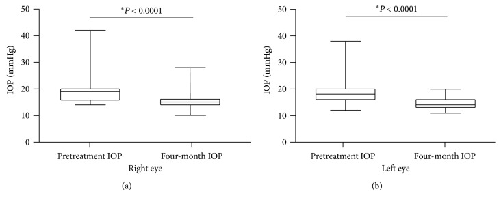
Results: Seventy-eight eyes of 46 patients diagnosed with POAG were studied: 58 eyes with maximum tolerated medical treatment and 20 eyes without treatment (naïve). The mean baseline IOP on the treated POAG group was 19.25 mmHg ± 4.71. Baseline IOP on the naïve group was 20.38 mmHg ± 3.28. At the four-month follow-up visit, the mean IOP value on the treatment group was 14.41 mmHg ± 2.06 (P < 0.0001). The obtained mean IOP measurement on the treatment-naïve group was 15.29 mmHg ± 2.28 (P < 0.0001).

Conclusions: The hypotensive response obtained using transpalpebral electrical stimulation on POAG patients, both on treatment-naïve patients and on patients receiving maximum tolerable treatment, was statistically significant when comparing basal IOP measurements to those obtained at the four-month follow-up visit.


Figures

Figure 1
Figure 1
Transpalpebral electrical microstimulation procedure on a POAG patient.
Figure 2
Figure 2
(a) Biphasic waveform applied on the most reported TES; (b) a new voltage waveform approach used in TPES.
Figure 3
Figure 3
(a) Comparison between IOP values obtained before electrical stimulation to those measured at the four-month follow-up visit for POAG patients receiving the maximum tolerated medical treatment. (b) Comparison between IOP values obtained before electrical stimulation to those measured at the four-month follow-up visit from naïve patients. Wilcoxon matched-pairs signed rank test.
Figure 4
Figure 4
IOP measurements obtained from POAG patients without treatment (naïve): (a) comparison of IOP values before electrical stimulation to those obtained at the four-month follow-up visit for the right eye; (b) comparison of IOP values before electrical stimulation to those obtained at the four-month follow-up visit in the left eye. Wilcoxon matched-pairs signed rank test.
Figure 5
Figure 5
IOP measurements obtained from POAG patients receiving maximum tolerated medical treatment: (a) comparison between IOP values obtained before electrical stimulation to those obtained at the four-month follow-up visit for the right eye; (b) comparison between IOP values obtained before electrical stimulation to those obtained at the four-month follow-up visit for the left eye. Wilcoxon matched-pairs signed rank test.
Figure 6
Figure 6
Coefficient of correlation (r) and linear regression analysis (R2) between POAG patients' years of age and IOP measurement at the four-month follow-up visit. Pearson correlation coefficients (r), 95% CI, and two-tailed P value.

Similar articles

Cited by

References

    1. Tham Y. C., Li X., Wong T. Y., Quigley H. A., Aung T., Cheng C. Y. Global prevalence of glaucoma and projections of glaucoma burden through 2040: a systematic review and meta-analysis. Ophthalmology. 2014;121(11):2081–2090. doi: 10.1016/j.ophtha.2014.05.013. - DOI - PubMed
    1. Schmidl D., Schmetterer L., Garhöfer G., Popa-Cherecheanu A. Pharmacotherapy of glaucoma. Journal of Ocular Pharmacology and Therapeutics. 2015;31(2):63–77. doi: 10.1089/jop.2014.0067. - DOI - PMC - PubMed
    1. Tezel G., Yang X., Luo C., et al. Oxidative stress and the regulation of complement activation in human glaucoma. Investigative Opthalmology and Visual Science. 2010;51(10):5071–5082. doi: 10.1167/iovs.10-5289. - DOI - PMC - PubMed
    1. Harris A., Siesky B., Wirostko B. Cerebral blood flow in glaucoma patients. Journal of Glaucoma. 2013;22:S46–S48. doi: 10.1097/ijg.0b013e3182934b6b. - DOI - PMC - PubMed
    1. Saccà S. C., Pulliero A., Izzotti A. The dysfunction of the trabecular meshwork during glaucoma course. Journal of Cellular Physiology. 2015;230(3):510–525. doi: 10.1002/jcp.24826. - DOI - PubMed

LinkOut - more resources