. 2018 Jul 11:2018:8461380.
doi: 10.1155/2018/8461380. eCollection 2018.

A Simple Separation Method of the Protein and Polystyrene Bead-Labeled Protein for Enhancing the Performance of Fluorescent Sensor

Affiliations

A Simple Separation Method of the Protein and Polystyrene Bead-Labeled Protein for Enhancing the Performance of Fluorescent Sensor

Hye Jin Kim et al. J Anal Methods Chem. .

Abstract

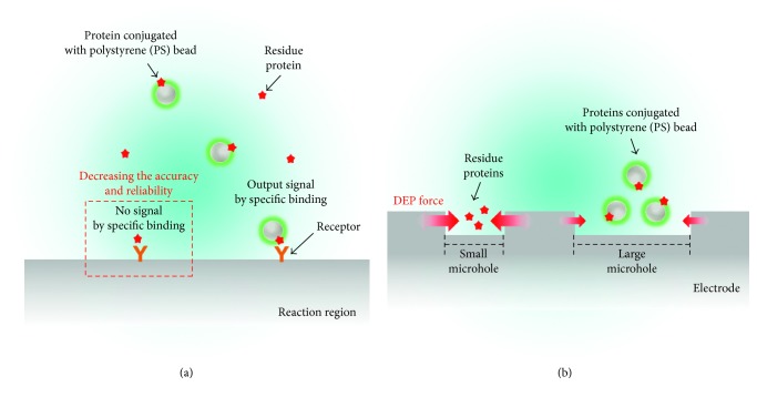
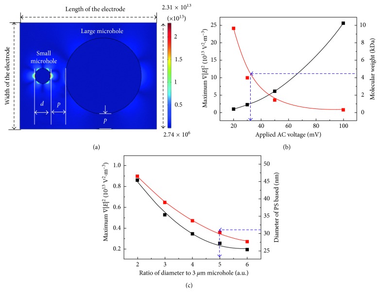
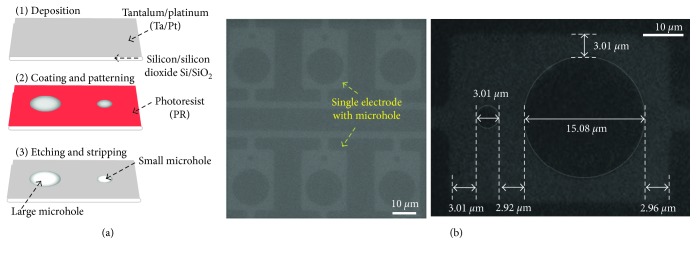
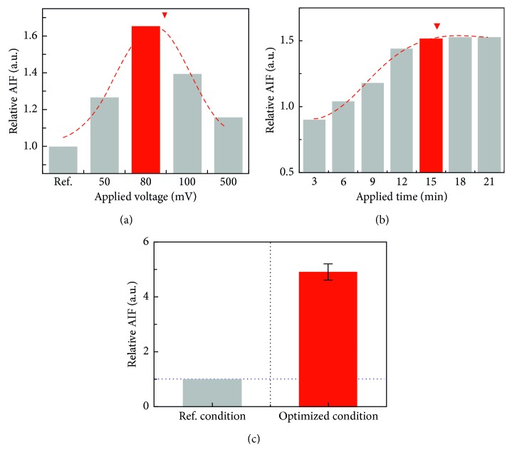
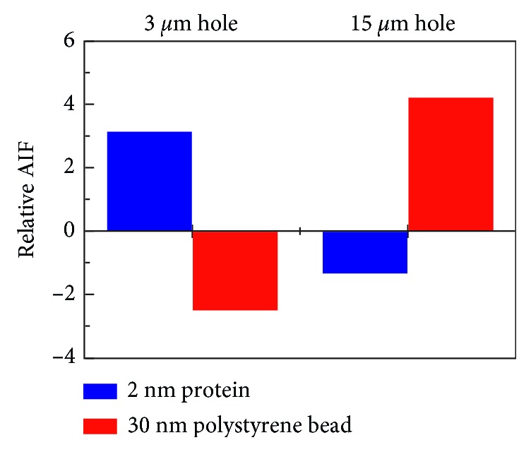
Dielectrophoresis- (DEP-) based separation method between a protein, amyloid beta 42, and polystyrene (PS) beads in different microholes was demonstrated for enhancement of performance for bead-based fluorescent sensor. An intensity of ∇|E|2 was relative to a diameter of a microhole, and the diameters of two microholes for separation between the protein and PS beads were simulated to 3 μm and 15 μm, respectively. The microholes were fabricated by microelectromechanical systems (MEMS). The separation between the protein and the PS beads was demonstrated by comparing the average intensity of fluorescence (AIF) by each molecule. Relative AIF was measured in various applying voltage and time conditions, and the conditions for allocating the PS beads into 15 μm hole were optimized at 80 mV and 15 min, respectively. In the optimized condition, the relative AIF was observed approximately 4.908 ± 0.299. Finally, in 3 μm and 15 μm hole, the AIFs were approximately 3.143 and -1.346 by 2 nm of protein and about -2.515 and 4.211 by 30 nm of the PS beads, respectively. The results showed that 2 nm of the protein and 30 nm of PS beads were separated by DEP force in each microhole effectively, and that our method is applicable as a new method to verify an efficiency of the labeling for bead-based fluorescent sensor ∇|E|2.


Figures

Figure 1
Figure 1
Schematic illustration of a simple separation method of the protein and protein conjugated with polystyrene (PS) beads. (a) Intensity of fluorescence by a specific binding of the protein conjugated with PS beads decreased due to a specific binding of the nonconjugated protein, expressed as residue protein. (b) Residue protein and protein conjugated with PS beads were separated by the dielectrophoresis (DEP) force in different microholes, respectively.
Figure 2
Figure 2
Simulation for separating the molecules by DEP force in the two microholes. (a) Distribution of ∇|E|2 at the top view of the electrode. The diameter of the small microhole and pitch between two microholes were represented as “d” and “p,” respectively. (b) According to the applied voltage, maximum intensity of ∇|E|2 occurring in the 3 μm hole increased, whereas molecular weight of the protein, allocated into the microhole, decreased. (c) Maximum intensity of ∇|E|2 in the other microhole and the diameter of PS beads, allocated into the hole, decreased according to the increase of the diameter.
Figure 3
Figure 3
Fabrication of the single electrode consisting of two different-sized microholes by MEMS technology. (a) Schematic illustration of the fabrication process of the microholes. (b) Microscopic image of the two different microholes.
Figure 4
Figure 4
Optimization of the DEP condition by measuring the average intensity of fluorescence (AIF) of the PS beads in the 15 μm hole. Relative AIF was verified according to (a) the applied voltage and (b) the applied time of AC voltage. (c) Relative AIF by the PS beads in the 15 μm hole was compared in each reference and optimized DEP condition.
Figure 5
Figure 5
Relative AIF by 2 nm of the protein and 30 nm of the PS beads in 3 μm and 15 μm holes, respectively.

Similar articles

References

    1. Chen X., Smith L. M., Bradbury E. M. Site-specific mass tagging with stable isotopes in proteins for accurate and efficient protein identification. Analytical Chemistry. 2000;72(6):1134–1143. doi: 10.1021/ac9911600. - DOI - PubMed
    1. Lummer M., Humpert F., Wiedenlübbert M., Sauer M., Schüttpelz M., Staiger D. A new set of reversibly photoswitchable fluorescent proteins for use in transgenic plants. Molecular Plant. 2013;6(5):1518–1530. doi: 10.1093/mp/sst040. - DOI - PubMed
    1. Sahoo H. Fluorescent labeling techniques in biomolecules: a flashback. RSC Advances. 2012;2(18):7017–7029. doi: 10.1039/c2ra20389h. - DOI
    1. Jung D., Min K., Jung J., Jang W., Kwon Y. Chemical biology-based approaches on fluorescent labeling of proteins in live cells. Molecular BioSystems. 2013;9(5):862–872. doi: 10.1039/c2mb25422k. - DOI - PubMed
    1. Bonar M. M., Tilton J. C. High sensitivity detection and sorting of infectious human immunodeficiency virus (HIV-1) particles by flow virometry. Virology. 2017;505:80–90. doi: 10.1016/j.virol.2017.02.016. - DOI - PMC - PubMed

LinkOut - more resources