Review
. 2018 Oct;10(5):1337-1348.
doi: 10.1007/s12551-018-0448-x. Epub 2018 Aug 16.

Cholesterol-dependent cytolysins: from water-soluble state to membrane pore

Affiliations
Review

Cholesterol-dependent cytolysins: from water-soluble state to membrane pore

Michelle P Christie et al. Biophys Rev. 2018 Oct.

Abstract

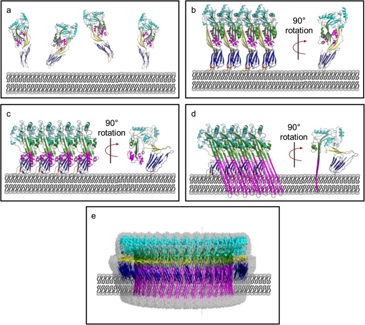
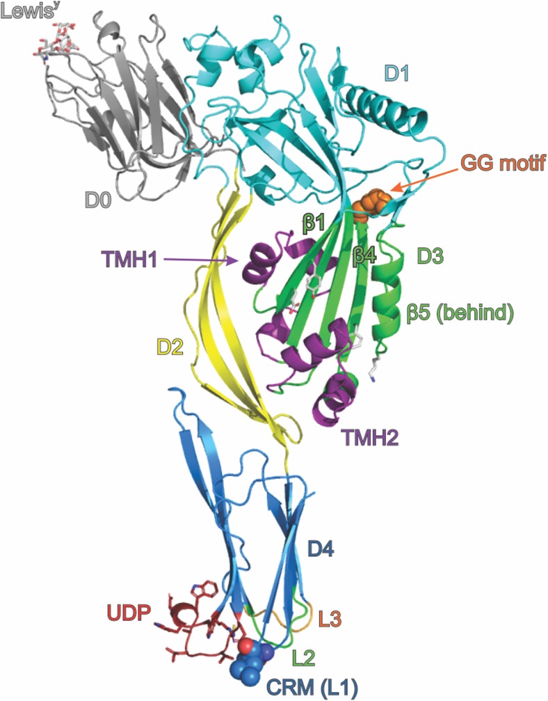
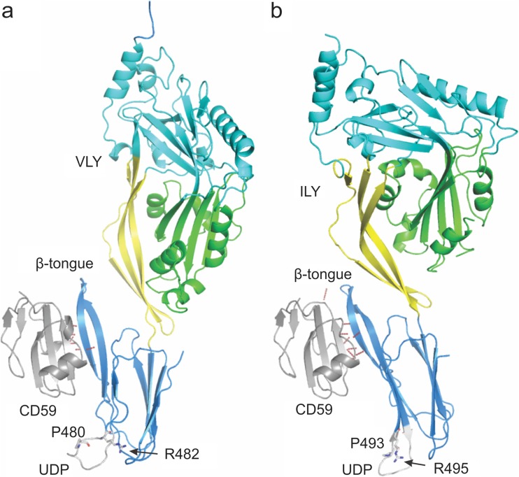
The cholesterol-dependent cytolysins (CDCs) are a family of bacterial toxins that are important virulence factors for a number of pathogenic Gram-positive bacterial species. CDCs are secreted as soluble, stable monomeric proteins that bind specifically to cholesterol-rich cell membranes, where they assemble into well-defined ring-shaped complexes of around 40 monomers. The complex then undergoes a concerted structural change, driving a large pore through the membrane, potentially lysing the target cell. Understanding the details of this process as the protein transitions from a discrete monomer to a complex, membrane-spanning protein machine is an ongoing challenge. While many of the details have been revealed, there are still questions that remain unanswered. In this review, we present an overview of some of the key features of the structure and function of the CDCs, including the structure of the secreted monomers, the process of interaction with target membranes, and the transition from bound monomers to complete pores. Future directions in CDC research and the potential of CDCs as research tools will also be discussed.

Keywords: Cholesterol-binding protein; Cholesterol-dependent cytolysin; Membrane-protein interactions; Pore-forming toxin.


Conflict of interest statement

Conflict of interest

M.P. Christie declares that she has no conflict of interest. B.A. Johnstone declares that she has no conflict of interest. R.K. Tweten declares that he has no conflict of interest. M.W. Parker declares that he has no conflict of interest. C.J. Morton declares that he has no conflict of interest.

Ethical approval

This article does not contain any studies with human participants or animals performed by any of the authors.

Figures

Fig. 1
Fig. 1
CDCs are secreted as soluble monomers (a), which bind to cholesterol on the surface of target cell membranes and oligomerise to form an early prepore structure (b). This involves slight insertion of the undecapeptide (UDP) loops in D4 into the lipid bilayer. CDC molecules in the early pre-pore undergo a 40 Å vertical collapse in D2, following disruption of the D3 interface (c). Full membrane insertion requires each monomer to contribute two TMH regions, which unfurl and insert in the membrane as β-hairpins (d). These β-hairpins form the β-barrel pore, which is typically about 300 Å in diameter and consists of between 30 and 50 individual monomers (e). All figures generated in Pymol (Schrodinger 2015). a, b Generated in Pymol using PDB: 5AOD. c is a model generated from PDB: 5AOD and PDB: 5LY6. d, c Generated using PDB 5LY6, the fitted model for the cryo-EM structure of the PLY pore complex
Fig. 2
Fig. 2
Diagrammatic representation of the structure of a CDC based on the structure of PFO (PDB: 1PFO) and D0 from LLY complexed with LewisY (PDB: 4GWI). The protein is shown as a cartoon, coloured by domain, with D0 grey, D1 cyan, D2 yellow, D3 green and D4 blue. D0, present in LLY, is shown with LewisY occupying the lectin-binding site. Key features of the structure are indicated, including the undecapeptide (UDP, shown as sticks), the cholesterol recognition motif (CRM, shown as spheres), the di-glycine motif that provides a pivot point during oligomerisation and pore formation (shown as orange spheres) and D3 residues mentioned in the text Y181, E183, F318 and K336 (shown as white sticks, unlabelled). The two transmembrane hairpins TMH1 and TMH2 are shown in their helical bundle state, coloured purple. Other labelled features are β-strands β1, β4 and β5 (discussed in the text) and the L2 and L3 loops of D4
Fig. 3
Fig. 3
Diagrammatic representation of the structures of the a VLY-CD59 and b ILY-CD59 complexes. The proteins are shown as cartoons coloured by domain with D1 cyan, D2 yellow, D3 green and D4 blue for the CDC and CD59 in grey. Hydrogen bonds identified between CD59 and the associated CDC are shown as dashed red lines. The β-tongue, UDP, conserved UDP arginine and unusual UDP proline are labelled. The structures were oriented after being superimposed on the CD59 molecules to emphasise the position of the D4 β-tongue interaction with CD59

References

    1. Bavdek A, et al. Sterol and pH interdependence in the binding, oligomerization, and pore formation of listeriolysin O. Biochemistry. 2007;46:4425–4437. doi: 10.1021/bi602497g. - DOI - PubMed
    1. Bhakdi S, Tranum-Jensen J, Sziegoleit A. Mechanism of membrane damage by streptolysin-O. Infect Immun. 1985;47:52–60. - PMC - PubMed
    1. Bonev BB, Gilbert RJ, Andrew PW, Byron O, Watts A. Structural analysis of the protein/lipid complexes associated with pore formation by the bacterial toxin pneumolysin. J Biol Chem. 2001;276:5714–5719. doi: 10.1074/jbc.M005126200. - DOI - PubMed
    1. Bourdeau RW, et al. Cellular functions and X-ray structure of anthrolysin O, a cholesterol-dependent cytolysin secreted by Bacillus anthracis. J Biol Chem. 2009;284:14645–14656. doi: 10.1074/jbc.M807631200. - DOI - PMC - PubMed
    1. Choe S, Bennett MJ, Fujii G, Curmi PM, Kantardjieff KA, Collier RJ, Eisenberg D. The crystal structure of diphtheria toxin. Nature. 1992;357:216–222. doi: 10.1038/357216a0. - DOI - PubMed

LinkOut - more resources