Review
. 2019 Jan;248(1):65-77.
doi: 10.1002/dvdy.24665. Epub 2018 Sep 12.

Compartmentalized Devices as Tools for Investigation of Human Brain Network Dynamics

Affiliations
Review

Compartmentalized Devices as Tools for Investigation of Human Brain Network Dynamics

Joseph A Fantuzzo et al. Dev Dyn. 2019 Jan.

Abstract

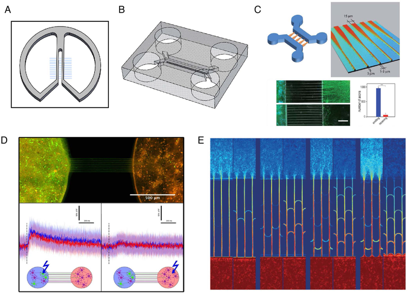
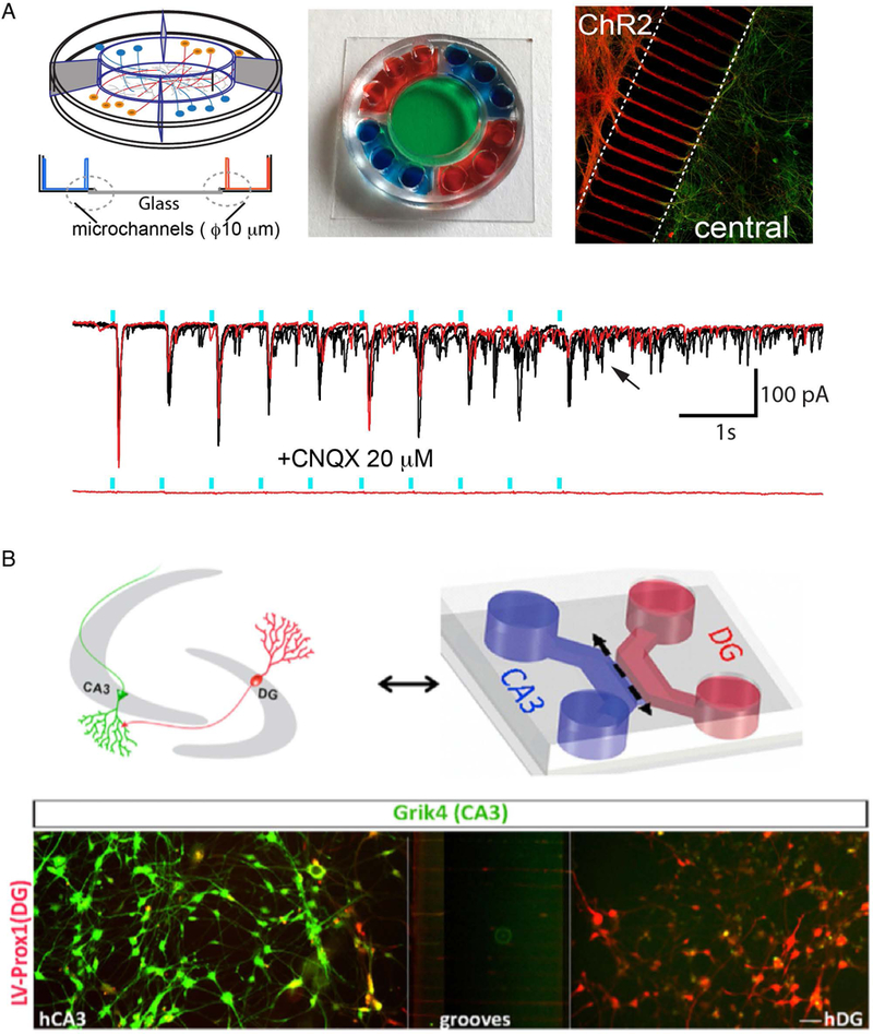
Neuropsychiatric disorders have traditionally been difficult to study due to the complexity of the human brain and limited availability of human tissue. Induced pluripotent stem (iPS) cells provide a promising avenue to further our understanding of human disease mechanisms, but traditional 2D cell cultures can only provide a limited view of the neural circuits. To better model complex brain neurocircuitry, compartmentalized culturing systems and 3D organoids have been developed. Early compartmentalized devices demonstrated how neuronal cell bodies can be isolated both physically and chemically from neurites. Soft lithographic approaches have advanced this approach and offer the tools to construct novel model platforms, enabling circuit-level studies of disease, which can accelerate mechanistic studies and drug candidate screening. In this review, we describe some of the common technologies used to develop such systems and discuss how these lithographic techniques have been used to advance our understanding of neuropsychiatric disease. Finally, we address other in vitro model platforms such as 3D culture systems and organoids and compare these models with compartmentalized models. We ask important questions regarding how we can further harness iPS cells in these engineered culture systems for the development of improved in vitro models. Developmental Dynamics 248:65-77, 2019. © 2018 Wiley Periodicals, Inc.

Keywords: 3D culture; Huntington's Disease; axonal transport; compartmentalization; induced neurons; microfluidics; neurocircuitry; neuropsychiatric disorders; organoids; stem cells.


Figures

Figure 1.
Figure 1.
Tools for compartmentalized systems. A) Campenot chamber (re-drawn from Zweifel et al. (Zweifel et al., 2005) and Millet and Gillette (Millet and Gillette, 2012)). Scratches in collagen surface produce channels which direct axonal outgrowth. B) Compartmentalized device made from PDMS (re-drawn from Taylor et al. (Taylor et al., 2005)). Microchannels (3 μm tall, 10 μm wide) connect two opposing chambers. C) Axon diodes drive axonal outgrowth primarily in one direction. Wider channels (15 μm) provide greater opportunity for neurites to enter whereas thinner channels (3 μm) limit neurite entry. (Reproduced from Peyrin et al. (Peyrin et al., 2011) with slight modifications with permission.) D) Incorporation of optogenetics and calcium imaging in a culture system, coupled with axon diodes. Calcium imaging bursts in response to light seen in both compartments only when Channelrhodopsin-2-transduced neurons were stimulated. (Reproduced with permission under CCBY4.0(https://creativecommons.org/licenses/by/4.0/legalcode) from Renault et al. (Renault et al., 2015)with slight modifications.) E) “Return-to-sender” approach provides directionality between compartments. Axons entering from the “receiver” side encounter bifurcations (arches) which return them back to their original chamber. Axons from the “sender” side do not encounter bifurcations and pass through to the “receiver” side (Reproduced from Renault et al. (Renault et al., 2016) with permission.)
Figure 2.
Figure 2.
Examples of compartmentalization for modeling Huntington’s Disease. A) Zhao et al. used twocompartment devices for investigating the role of mHTT and rescuing effects of TriC. Tau and DARPP32 staining verify the presence of neurons on both sides but DARPP32-positive neurons restricted to one compartment. (Reproduced from Zhao et al. (Zhao et al., 2016) with permission). B) Three-compartment device developed by Virlogeux et al. A specific cortical-to-striatal setup was achieved through extending microchannels greater than 450 μm and creating a laminin gradient in the striatal chamber. (Reproduced from Virlogeux et al. (Virlogeux et al., 2018) with permission).
Figure 3.
Figure 3.
Devices using human induced neurons. A) Five-compartment device developed by Fantuzzo et al. (Fantuzzo et al., 2017). Center chamber enables access for electrophysiological analysis of circuit. Optogenetic stimulation (blue bar) is coupled to post-synaptic currents, indicating formation of a functional circuit. (Reproduced from Fantuzzo et al. (Fantuzzo et al., 2017) with permission). B) Hippocampal circuit (CA3-DG) modeled with human neurons. (Reproduced from Sarkar et al. (Sarkar et al., 2018) with permission).
Figure 4.
Figure 4.
Alternative approaches for modeling neuropsychiatric disorders with human cells. A) Layered hydrogel model used for the study of Rett syndrome. (Reproduced from Zhang et al. (Zhang et al., 2016) with permission). B) A 3D culture in Matrigel hydrogel produced through 3D-printed apparatus. (Reproduced from Alessandri et al. (Alessandri et al., 2016) with permission). C) The OrganoPlate®, which provides a high-throughput 3D culture model for human induced neurons. (Reproduced under CCBY4.0 (https://creativecommons.org/licenses/by/4.0/legalcode) from Wevers et al. (Wevers et al., 2016)). D) Generation of human organoids in Spin_ bioreactors by the Ming laboratory to study Zika virus. (Reproduced from Qian et al. (Qian et al., 2016) with permission).

References

    1. Alessandri K, Feyeux M, Gurchenkov B, Delgado C, Trushko A, Krause KH, Vignjevic D, Nassoy P, Roux A. 2016. A 3D printed microfluidic device for production of functionalized hydrogel microcapsules for culture and differentiation of human Neuronal Stem Cells (hNSC). Lab Chip 16:1593–1604. - PubMed
    1. Atwal JK, Massie B, Miller FD, Kaplan DR. 2000. The TrkB-Shc Site Signals Neuronal Survival and Local Axon Growth via MEK and PI3-Kinase. Neuron 27:265–277. - PubMed
    1. Bailey A, Le Couteur A, Gottesman I, Bolton P, Simonoff E, Yuzda E, Rutter M. 2009. Autism as a strongly genetic disorder: evidence from a British twin study. Psychological Medicine 25:63. - PubMed
    1. Berdichevsky Y, Sabolek H, Levine JB, Staley KJ, Yarmush ML. 2009. Microfluidics and multielectrode array-compatible organotypic slice culture method. Journal of neuroscience methods 178:59–64. - PMC - PubMed
    1. Bernstein JG, Boyden ES. 2011. Optogenetic tools for analyzing the neural circuits of behavior. Trends Cogn Sci 15:592–600. - PMC - PubMed

Publication types

LinkOut - more resources