

Metastatic thymoma in the liver of a dog
- PMID: 30117785
- PMCID: PMC6505795
- DOI: 10.1177/1040638718791222
Metastatic thymoma in the liver of a dog
Abstract
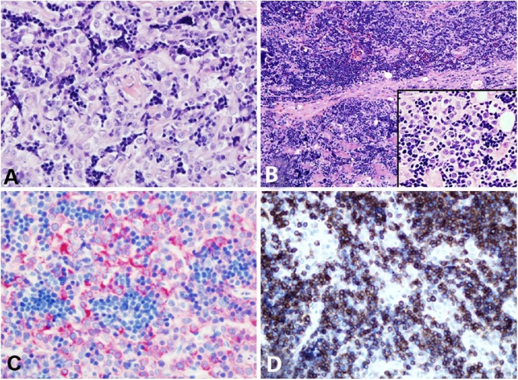
A 12-y-old neutered male Portuguese Water dog was presented because of a 1-y history of persistent hyporexia, diarrhea, and recurrent pyelonephritis. Abdominal ultrasound revealed hepatic nodules and diffuse splenomegaly, and radiographs revealed a mediastinal mass. Fine-needle aspirates of the liver, spleen, and mediastinal mass were suspicious for lymphoma. Flow cytometry identified small T cells that co-expressed CD4 and CD8 at all sites, most suspicious for thymoma, but lymphoma could not be ruled out. PCR for antigen receptor rearrangements analysis identified polyclonal amplification of the T-cell receptor genes, more consistent with thymoma than lymphoma. Histopathology of the liver and thymic mass confirmed thymoma with hepatic metastasis.
Keywords: Dogs; flow cytometry; hepatic metastasis; metastatic thymoma.
Conflict of interest statement
Figures


Similar articles
-
Thymoma-associated lymphocytosis in a dog.Vet Clin Pathol. 2014 Dec;43(4):584-8. doi: 10.1111/vcp.12196. Epub 2014 Oct 8. Vet Clin Pathol. 2014. PMID: 25295998
-
Pathology in Practice. Metastatic lymphocyte-rich thymoma in a dog.J Am Vet Med Assoc. 2017 Feb 15;250(4):387-390. doi: 10.2460/javma.250.4.387. J Am Vet Med Assoc. 2017. PMID: 28165310 No abstract available.
-
CD4 and CD8 double-negative immunophenotype of thymoma-associated lymphocytes in a dog.J Vet Diagn Invest. 2020 Nov;32(6):918-922. doi: 10.1177/1040638720948628. Epub 2020 Aug 19. J Vet Diagn Invest. 2020. PMID: 32814519 Free PMC article.
-
Thymoma: current diagnosis and treatment.Chin Med J (Engl). 2013;126(11):2186-91. Chin Med J (Engl). 2013. PMID: 23769581
-
Ectopic Cervical Thymoma in a Patient Diagnosed With Graves Disease: A Systematic Literature Review.J Clin Endocrinol Metab. 2024 Apr 19;109(5):1198-1201. doi: 10.1210/clinem/dgad635. J Clin Endocrinol Metab. 2024. PMID: 37897424
Cited by
-
Canine Epithelial Thymic Tumors: Outcome in 28 Dogs Treated by Surgery.Animals (Basel). 2021 Dec 2;11(12):3444. doi: 10.3390/ani11123444. Animals (Basel). 2021. PMID: 34944221 Free PMC article.
References
-
- Al-Zubaidy AJ. Malignant thymoma with metastases in a dog. Vet Rec 1981;109:490–492. - PubMed
-
- Atwater SW, et al. Thymoma in dogs: 23 cases (1980–1991). J Am Vet Med Assoc 1994;205:1007–1013. - PubMed
-
- Batlivala TP, et al. Paraneoplastic T cell lymphocytosis associated with a thymoma in a dog. J Small Anim Pract 2010;51:491–494. - PubMed
-
- Bismarck D,et al. . Canine CD4+CD8+ double positive T cells in peripheral blood have features of activated T cells. Vet Immunol Immunopathol 2012;149:157–166. - PubMed
Publication types
MeSH terms
Grants and funding
LinkOut - more resources
Full Text Sources
Other Literature Sources
Medical
Research Materials
