. 2018 Oct 1;50(10):862-875.
doi: 10.1152/physiolgenomics.00076.2018. Epub 2018 Aug 17.

Effects of placental growth factor deficiency on behavior, neuroanatomy, and cerebrovasculature of mice

Affiliations

Effects of placental growth factor deficiency on behavior, neuroanatomy, and cerebrovasculature of mice

Vanessa R Kay et al. Physiol Genomics. .

Abstract

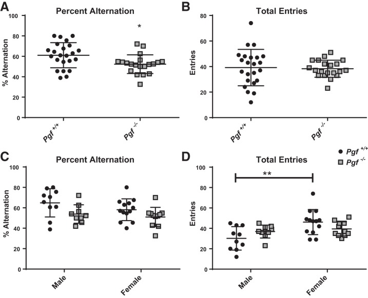
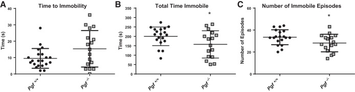
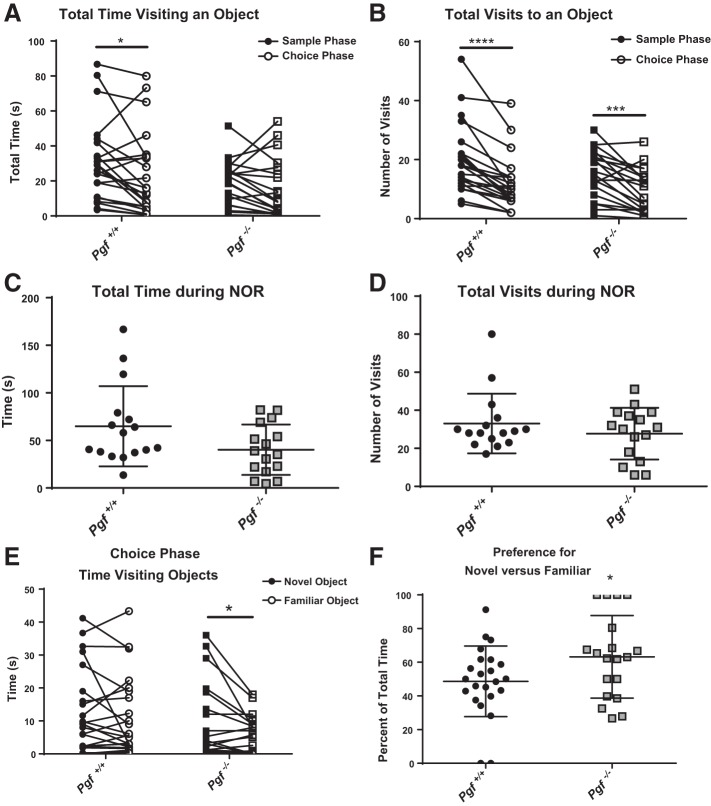
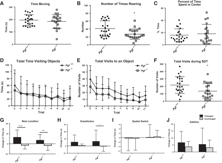
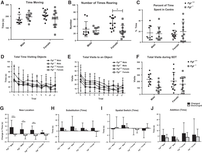
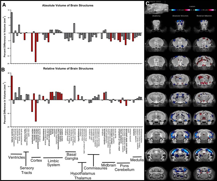
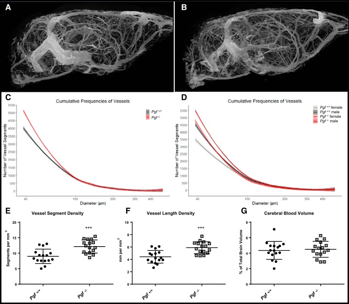
Preeclampsia, a hypertensive syndrome occurring in 3-5% of human pregnancies, has lifelong health consequences for fetuses. Cognitive ability throughout life is altered, and adult stroke risk is increased. One potential etiological factor for altered brain development is low concentrations of proangiogenic placental growth factor (PGF). Impaired PGF production may promote an antiangiogenic fetal environment during neural and cerebrovascular development. We previously reported delayed vascularization of the hindbrain, altered retinal vascular organization, and less connectivity in the circle of Willis in Pgf-/- mice. We hypothesized Pgf-/- mice would have impaired cognition and altered brain neuroanatomy in addition to compromised cerebrovasculature. Cognitive behavior was assessed in adult Pgf-/- and Pgf+/+ mice by four paradigms followed by postmortem high-resolution MRI of neuroanatomy. X-ray microcomputed tomography imaging investigated the three-dimensional cerebrovascular geometry in another cohort. Pgf-/- mice exhibited poorer spatial memory, less depressive-like behavior, and superior recognition of novel objects. Significantly smaller volumes of 10 structures were detected in the Pgf-/- compared with Pgf+/+ brain. Pgf-/- brain had more total blood vessel segments in the small-diameter range. Lack of PGF altered cognitive functions, brain neuroanatomy, and cerebrovasculature in mice. Pgf-/- mice may be a preclinical model for the offspring effects of low-PGF preeclampsia gestation.

Keywords: brain development; magnetic resonance imaging; microcomputed tomography imaging; preeclampsia; pregnancy.


Figures

Fig. 1.
Fig. 1.
Spatial learning in Pgf−/− mice. Compared with Pgf+/+ mice, Pgf−/− mice exhibited a significantly lower percent alternation (A) despite no difference in the total number of arm entries (B) in the Y-maze spontaneous alternation test (YMSAT). When stratified by sex, there was no significant difference in percent alternation (C), although female Pgf+/+ mice made significantly more entries than Pgf+/+males (D). Each mouse completed the 10 min YMSAT once. Data were analyzed with unpaired, two-tailed t-tests. Graphs show means ± SD. Black circles represent Pgf+/+ mice, while gray squares represent Pgf−/− mice; n = 23 Pgf+/+ and 20 Pgf−/− male and female mice. *P < 0.05.
Fig. 2.
Fig. 2.
Depressive-like behavior in Pgf−/− mice. Compared with Pgf+/+ mice, Pgf−/− mice showed no difference in time to immobility (A), although they spent less time immobile (B) and had fewer immobile episodes (C) on the tail suspension test (TST). Each mouse completed the 6 min TST once. Unpaired two-tailed t-tests or Mann-Whitney tests for data that were not normally distributed were used for analysis. Graphs show means ± SD. Black circles represent Pgf+/+ mice, while gray squares represent Pgf−/− mice; n = 23 Pgf+/+ and 20 Pgf−/− male and female mice. *P < 0.05.
Fig. 3.
Fig. 3.
Pgf−/− mouse performance on the novel object recognition (NOR) test. On the NOR test, Pgf+/+ but not Pgf−/− mice spent less time exploring the objects during the choice phase compared with the sample phase, although there was no difference between genotypes in either phase (A). Both Pgf+/+ and Pgf−/− mice made fewer visits to the objects during the choice phase compared with the sample phase with no significant difference between genotypes in either phase (B). There was no significant difference in time spent exploring objects (C) or number of visits to the objects (D) over the entire NOR. In the choice phase, Pgf−/− mice were able to differentiate between the novel object and familiar objects and spent more time exploring the novel object (E). Correspondingly, Pgf−/− mice exhibited greater preference for the novel object compared with Pgf+/+ mice as measured by the percent of total time spent exploring the novel object (F). Each mouse completed three 10 min trials. In the first trial, the arena was empty. In the second trial, two identical objects were present for the sample phase. In the third trial, one familiar object and one novel object were present for the choice phase. Data were analyzed with repeated-measures two-way ANOVA, unpaired, two-tailed t-tests, or Mann-Whitney tests as appropriate. Graphs show values for individual mice in the sample and choice phases (A, B, E), or means ± SD (C, D, F). Circles represent Pgf+/+ mice, while squares represent Pgf−/− mice; n = 23 Pgf+/+ and 20 Pgf−/− male and female mice. *P < 0.05, ***P < 0.001, ****P < 0.0001.
Fig. 4.
Fig. 4.
Pgf−/− mouse performance on the serial dishabituation test (SDT). In the first trial of the SDT, Pgf+/+ and Pgf−/− mice spent similar amounts of time moving (A), although Pgf−/− mice reared fewer times (B). The time spent in the center of the arena as a percentage of total time was also similar between Pgf+/+ and Pgf−/− mice (C). Pgf+/+ and Pgf−/− mice spent similar amounts of time exploring the objects in each trial (D), although Pgf−/− mice tended to make fewer visits to the objects (E). Pgf−/− mice had a lower total number of visits over the entire SDT (F). Pgf+/+ and Pgf−/− mice exhibited a significantly greater increase in time investigating the changed object compared with the unchanged objects during the New Location trial (G). Only Pgf−/− mice spent significantly greater time investigating the changed object in the Substitution trial (H). Neither the Pgf+/+or Pgf−/− had significantly greater increases in exploration of the changed objects during the Spatial Switch (I) or Addition (J) trials. Each mouse underwent 11 consecutive trials in the SDT. The first trial allowed accommodation to the empty arena and was used to analyze activity and anxiety-like behavior. The second to fourth trials familiarized the mice with an array of objects. The geometry of the array was distorted (New Location, Addition) or objects were exchanged without altering the geometry (Spatial Switch, Substitution) in later trials. Changes in the time spent investigating changed and unchanged objects relative to the baseline trial experienced directly previously are presented. Data were analyzed with unpaired, two-tailed t-tests, Mann-Whitney tests and two-way repeated-measures ANOVAs. Graphs show means ± SD. Circles represent Pgf+/+ mice, while squares represent Pgf−/− mice. Black bars show the change in time spent investigating the changed object(s), while light gray bars show the change in time investigating the unchanged objects; n = 23 Pgf+/+ and 20 Pgf−/− male and female mice. *P < 0.05, **P < 0.01, ****P < 0.0001.
Fig. 5.
Fig. 5.
Pgf−/− mouse performance on the SDT by sex. In the first trial of the SDT, there was no difference in time spent moving (A) between Pgf+/+ and Pgf−/− males and females, although Pgf+/+ females reared significantly more times than Pgf−/− females (B). There was also no difference in percent of time spent in the center between groups (C). Time spent exploring (D) and number of visits made (E) to the objects decreased over the trials but with no significant difference between groups. Likewise, total number of visits made to the objects over the entire SDT was not different between groups (F). Pgf+/+ males, Pgf+/+ females and Pgf−/− males spent significantly more time exploring the changed object than the unchanged objects in the New Location trial (G). However, none of the groups spent significantly more time exploring the changed object(s) relative to the unchanged objects in the Substitution (H), Spatial Switch (I), or Addition (J) trials. No clear sex difference in object recognition memory was apparent. Data were analyzed by two-way ANOVAs and linear mixed models as appropriate. Graphs show means ± SD. Circles represent Pgf+/+ mice, while squares represent Pgf−/− mice. Black bars show the change in time spent investigating the changed object(s), while light gray bars show the change in time investigating the unchanged objects; n = 23 Pgf+/+ and 20 Pgf−/− male and female mice. *P < 0.05, **P < 0.01, ***P < 0.001.
Fig. 6.
Fig. 6.
Brain structure volumes in Pgf−/− mice. MR structural analysis of mice that underwent behavioral testing (n = 8 males and females of each genotype with 1 Pgf−/− male excluded for poor image quality) revealed significant differences in the absolute volume of 10/62 brain structures between the Pgf+/+ and Pgf−/− mice (A). The difference in relative volume was significant in 14/62 areas with 4 areas smaller and 10 areas larger in the Pgf−/− mice (B). Graphs show percent difference with significance indicated by filled red bars. Voxel-wise comparison revealed size differences in widespread areas of the Pgf−/− brain seen in coronal sections (i–ix) of the MR scan (C). Significantly smaller (blue) absolute volume is complimented by significantly larger (red) relative volumes in other areas. Structures are identified as: abv, arbor vita of cerebellum; Amg, amygdala; BFB, basal forebrain; CbCx, cerebellar cortex; ml, corpus callosum; cp, cerebral peduncle; CPu, caudate/putamen; DG, dentate gyrus; EC, entorhinal cortex; fi, fimbria; FL, frontal lobe; fx, fornix; Hi, hippocampus; Hy, hypothalamus; icp, inferior cerebellar peduncle; LS, lateral septum; LV, lateral ventricle; MB, midbrain; mtt, mammillothalamic tract; Me, medulla, ml, medial lemniscus; MS, medial septum; NA, nucleus accumbens; OB, olfactory bulb; OL, occipital lobe; pag, periaqueductal gray matter; Po, pons; PPS, pre-parasubiculum; PTL, parietotemporal lobe; sc, superior colliculus; scp, superior cerebellar peduncle; sm, stria medullaris; Th, thalamus; TV, third ventricle. Data were analyzed with a false discovery rate of 0.1 for multiple comparisons.
Fig. 7.
Fig. 7.
Cerebrovasculature in Pgf−/− mice. Microcomputed tomography (µCT) imaging of the cerebral vasculature in Pgf+/+ (A) and Pgf−/− (B) mice (n = 8 males and 8 females of each genotype) revealed Pgf−/− mice had greater numbers of 40–100 µm diameter vessel segments in the brain (C). Both male and female Pgf−/− mice had more small-diameter vessels relative to Pgf+/+ controls (D). Vessel segment density (E) and vessel length density (F) were greater in Pgf−/− mice, but cerebral blood volume was not different (G). Cumulative frequency histograms were analyzed with fitted spline models and linear modeling of the spline coefficients. Vessel segment density, vessel length density, and cerebral blood volume were compared by unpaired two-tail t-tests. Graphs show averaged cumulative frequency histograms with 95% confidence interval or means ± SD. In the cumulative frequency histograms, Pgf+/+ mice are represented by gray and Pgf−/− mice represented by red with females and males are differentiated by darker and lighter shades, respectively. Black circles represent Pgf+/+, while gray squares represent Pgf−/− mice. ***P < 0.001.

Similar articles

Cited by

References

    1. Aasa KL, Zavan B, Luna RL, Wong PG, Ventura NM, Tse MY, Carmeliet P, Adams MA, Pang SC, Croy BA. Placental growth factor influences maternal cardiovascular adaptation to pregnancy in mice. Biol Reprod 92: 44, 2015. doi:10.1095/biolreprod.114.124677. - DOI - PMC - PubMed
    1. Barker DJ, Lampl M, Roseboom T, Winder N. Resource allocation in utero and health in later life. Placenta 33, Suppl 2: e30–e34, 2012. doi:10.1016/j.placenta.2012.06.009. - DOI - PubMed
    1. Beck H, Acker T, Püschel AW, Fujisawa H, Carmeliet P, Plate KH. Cell type-specific expression of neuropilins in an MCA-occlusion model in mice suggests a potential role in post-ischemic brain remodeling. J Neuropathol Exp Neurol 61: 339–350, 2002. doi:10.1093/jnen/61.4.339. - DOI - PubMed
    1. Benovská M, Opluštilová A, Pinkavová J, Hodická Z, Cermáková Z. The new possibilities in early diagnosis of preeclampsia by soluble fms-like tyrosine kinase-1 and placental growth factor in 16–20 weeks gestation. Lab Med 49: 112–117, 2017. doi:10.1093/labmed/lmx076. - DOI - PubMed
    1. Bergen NE, Bouwland-Both MI, Steegers-Theunissen RP, Hofman A, Russcher H, Lindemans J, Jaddoe VW, Steegers EA. Early pregnancy maternal and fetal angiogenic factors and fetal and childhood growth: the Generation R Study. Hum Reprod 30: 1302–1313, 2015. doi:10.1093/humrep/dev070. - DOI - PubMed

Publication types

LinkOut - more resources