. 2018 Aug 17;13(8):e0202263.
doi: 10.1371/journal.pone.0202263. eCollection 2018.

Trans-chalcone increases p53 activity via DNAJB1/HSP40 induction and CRM1 inhibition

Affiliations

Trans-chalcone increases p53 activity via DNAJB1/HSP40 induction and CRM1 inhibition

Gabriel Silva et al. PLoS One. .

Abstract

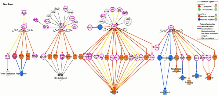
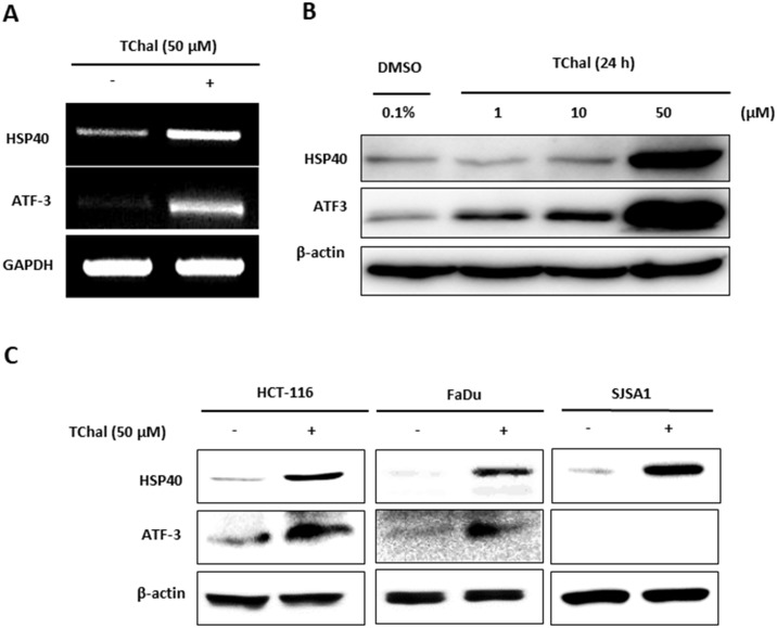
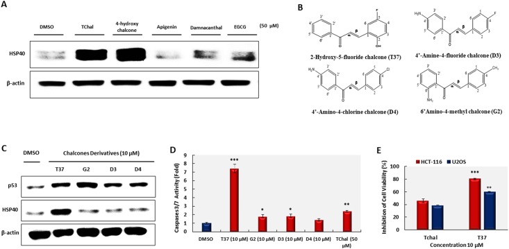
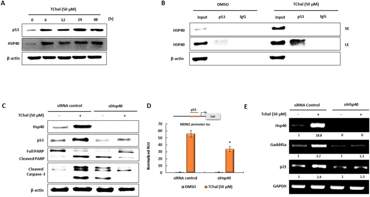
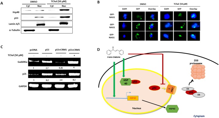
Naturally-occurring chalcones and synthetic chalcone analogues have been demonstrated to have many biological effects, including anti-inflammatory, anti-malarial, anti-fungal, and anti-oxidant/anti-cancerous activities. Compared to other chalcones, trans-chalcone exhibits superior inhibitory activity in cancer cell growth as shown via in vitro assays, and exerts anti-cancerous effects via the activation of the p53 tumor suppressor protein. Thus, characterization of the specific mechanisms, by which trans-chalcone activates p53, can aid development of new chemotherapeutic drugs that can be used individually or synergistically with other drugs. In this report, we found that trans-chalcone modulates many p53 target genes, HSP40 being the most induced gene in the RNA-Seq data using trans-chalcone-treated cells. CRM1 is also inhibited by trans-chalcone, resulting in the accumulation of p53 and other tumor suppressor proteins in the nucleus. Similar effects were seen using trans-chalcone derivatives. Overall, trans-chalcone could provide a strong foundation for the development of chalcone-based anti-cancer drugs.


Conflict of interest statement

No competing interests exist.

Figures

Fig 1
Fig 1. TChal activated the p53 signaling pathway at the transcriptional level.
U2OS cells were treated with 50 μM of DMSO or TChal for 24 h. The RNA was isolated, and then RNA-Seq analysis was performed. The Ingenuity Pathway Analysis (IPA) software was applied to analyze the differential gene expression data. The p53 signaling pathway was activated by TChal, and the downstream effects of p53 activation induced by TChal was determined using the tools available on the IPA.
Fig 2
Fig 2. TChal induced Hsp40 and ATF3 expression in cancer cells.
A. U2OS cells were treated with DMSO or TChal for 24 h. After RNA isolation, RT-PCR was performed using primers for Hsp40 and ATF3, as well as the housekeeping gene, GAPDH. B. Western blot analysis of Hsp40 and ATF3 protein expression after TChal treatment. U2OS cells were treated with TChal at the indicated doses, and then cell lysates were subjected to Western blot analysis. C. Protein expression of Hsp40 and ATF3 in different cancer cells. HCT-116 (colorectal cancer), FaDu (oral cancer), and SJSA1 (bone cancer) were treated with DMSO or 50 μM of TChal for 24 h. Cell lysates were then subjected to Western blot analysis. A representative gel from 3 independent experiments are shown.
Fig 3
Fig 3. Effect of chalcone derivatives on p53 and Hsp40 protein expression, cell growth, and caspase-3/7 activity.
A. Western blot analysis of HSP40 protein expression after treatment with 50 μM of different compounds for 24 h. A representative gel from 3 independent experiments are shown. B. Structure of chalcone derivatives. These figures were drawn using ChemDraw Ultra 8.0. C. Protein levels of p53 and HSP40 after treatment with chalcone derivatives. U2OS cells were treated with derivatives at the indicated doses for 24 h, and then cell lysates were subjected to Western blotting. A representative gel from 3 independent experiments are shown. D. U2OS cells were treated with TChal (50 μM) or chalcone derivatives (10 μM) for 24 h. Caspase-3/7 activity using 30 μg of cell lysate was measured using the Apo-ONE Homogenous Caspase-Glo 3/7 Assay kit. Values are expressed as mean ± SD of three replicates. *, P ≤ 0.05; **, P ≤ 0.01; and ***, P ≤ 0.001 versus DMSO-treated cells. E. U2OS cells were treated with DMSO (0.1%) or 10 μM of TChal and T37 for 24 h, and then cell growth was measured via MTS assay. Values are expressed as mean ± SD of four replicates. **, P ≤ 0.01 and ***, P ≤ 0.001 versus TChal-treated cells.
Fig 4
Fig 4. The induction of HSP40 by TChal contributes to the stabilization of p53 activity.
A. U2OS cells were treated with TChal at the indicated dose and times. The p53 and HSP40 expressions were measured by western blot analysis. B. U2OS cells were treated with 50 μM of TChal for 24 h, and cell lysates were isolated with a modified RIPA buffer. The cell lysates were immunoprecipitated with 2 μg of p53 or normal IgG antibodies using A/G PLUS-agarose, as described in the Materials and Methods section, and then subjected to western blot analysis using Hsp40 antibody. C. U2OS cells were transfected with 30 nM of Hsp40 or control siRNAs for 24 h, followed by treatment with 50 μM of TChal for 24 h. Then, cells lysates were subjected to western blot analysis. D. Vectors containing the MDM2-Luc promoter were co-transfected with Hsp40 or control siRNAs into U2OS cells, and cells were treated with 50 μM of TChal for 24 h. Luciferase activity was measured; y-axis represents RLU (relative luciferase unit). E. HSP40 silencing suppressed the effect of TChal on p53-dependent transcription. U2OS cells were transfected with Hsp40 or control siRNAs, followed by treatment with 50 μM of TChal for 24 h. Total RNA was isolated, and RT-PCR was performed as described in the Materials and Methods section. A representative gel from 3 independent experiments are shown.
Fig 5
Fig 5. TChal regulates the translocation of p53 via CRM1 downregulation.
A. Free-magnetic beads or beads bound with TChal were obtained as described in the Materials and Methods section, and incubated overnight at 4 °C with cell lysates from U2OS cells overexpressing CRM1. The beads were washed and collected, and then subjected to Western blot analysis. B. Cells transfected with 2.5 μg of Flag-CRM1 vector were treated with 50 μM of TChal for 24 h. The cells lysates were obtained and subjected to western blotting. Left, Western blot using Flag antibody; Right, Western blot using CRM1 antibody. C. U2OS cells were co-transfected with 1.25 μg of Flag-CRM1 and Flag-p53 vectors, followed by treatment with TChal for 24 h. Cell lysates were isolated using modified RIPA buffer, immunoprecipitated with 2 μg of CRM1 or normal IgG antibodies as described in the Materials and Methods section, and then subjected to Western blot analysis using Flag antibody. A representative gel from 3 independent experiments are shown.
Fig 6
Fig 6. TChal regulates p53 activity via CRM1 inhibition.
A. U2OS cells were co-transfected with 2 μg of Flag-CRM1 and 0.4 μg of Flag-p53 vectors, followed by treatment with 50 μM of TChal for 24 h. The cytoplasmic (Cyt) and nuclear (Nuc) fractions were obtained and subjected to western blot analysis using Flag antibodies. A representative gel from 3 independent experiments are shown. B. U2OS cells, transfected with 2.5 μg of GFP-tagged NAG-1 (WT) and treated with TChal for 24 h, were fixed and analyzed by fluorescence using the EVOS FL Auto Cell Imaging System. DAPI was used to stain the nuclei. Three independent fields are shown. C. U2OS cells were transfected with 0.4 μg of Flag-p53 vector, or co-transfected with 0.4 μg of Flag-p53 and 2 μg of Flag-CRM1 vectors, and then treated with 50 μM of TChal for 24 h. Total RNA was isolated, and RT-PCR was performed as described in the Materials and Methods section. A representative gel from 3 independent experiments are shown. D. A proposed model for trans-chalcone’s action on the stabilization, translocation, and activity of the p53 protein. Trans-chalcone increases the expression of HSP40 protein, which stabilizes the native conformation of the p53 protein. Trans-chalcone interacts with and decreases CRM1 expression, promoting the accumulation of p53 into the nucleus, and thus increasing the transcriptional activity of p53.

References

    1. Dickson J, Flores L, Stewart M, LeBlanc R, Pati HN, Lee M, et al. Synthesis and Cytotoxic Properties of Chalcones: An Interactive and Investigative Undergraduate Laboratory Project at the Interface of Chemistry and Biology. J Chem Educ. 2006;83(6):934 10.1021/ed083p934 - DOI
    1. Mahapatra DK, Bharti SK, Asati V. Anti-cancer chalcones: Structural and molecular target perspectives. Eur J Med Chem. 2015;98:69–114. 10.1016/j.ejmech.2015.05.004. - DOI - PubMed
    1. Silva G, Fachin AL, Beleboni RO, Franca SC, Marins M. In vitro action of flavonoids in the canine malignant histiocytic cell line DH82. Molecules (Basel, Switzerland). 2013;18(12):15448–63. Epub 2013/12/20. 10.3390/molecules181215448 . - DOI - PMC - PubMed
    1. Kandaswamy N, Raveendiran N. A REVIEW ON BIOLOGICAL POTENTIAL OF CHALCONE HYBRIDS. IAJPR. 2014;4(6):3011–22.
    1. Haupt Y, Maya R, Kazaz A, Oren M. Mdm2 promotes the rapid degradation of p53. Nature. 1997;387(6630):296–9. 10.1038/387296a0 . - DOI - PubMed

Publication types

MeSH terms