. 2018 Aug 16;71(4):637-648.e5.
doi: 10.1016/j.molcel.2018.07.010.

An Mtb-Human Protein-Protein Interaction Map Identifies a Switch between Host Antiviral and Antibacterial Responses

Affiliations

An Mtb-Human Protein-Protein Interaction Map Identifies a Switch between Host Antiviral and Antibacterial Responses

Bennett H Penn et al. Mol Cell. .

Abstract

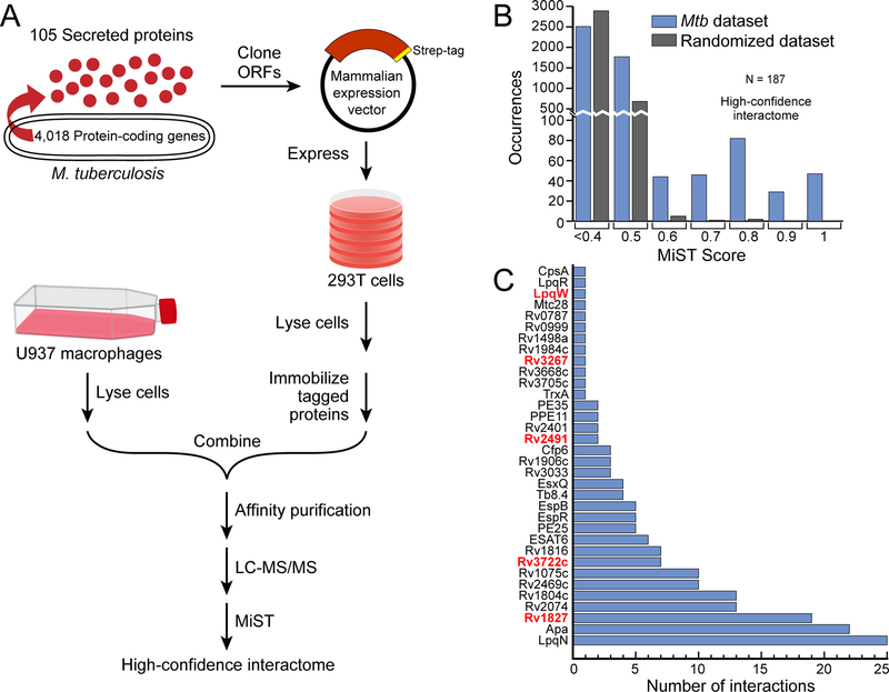
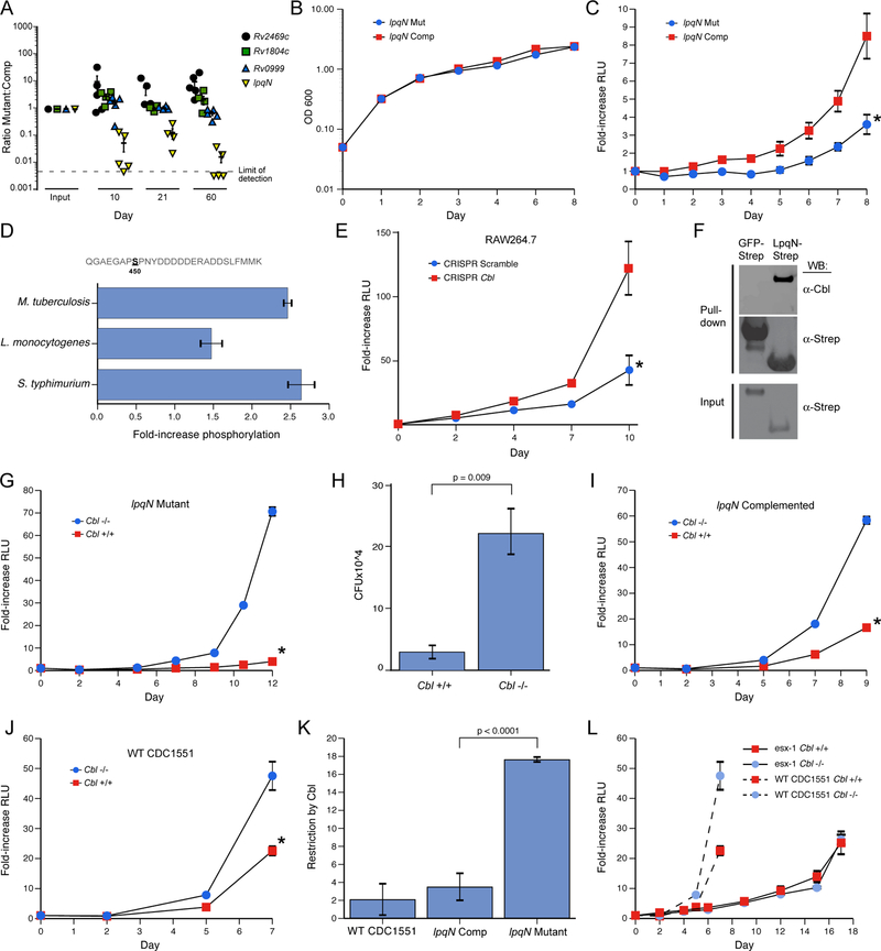
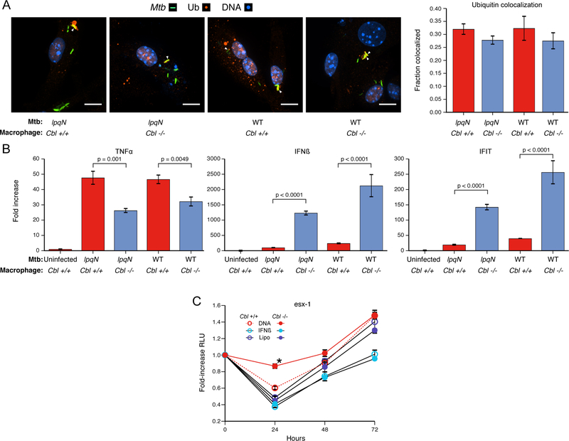
Although macrophages are armed with potent antibacterial functions, Mycobacterium tuberculosis (Mtb) replicates inside these innate immune cells. Determinants of macrophage intrinsic bacterial control, and the Mtb strategies to overcome them, are poorly understood. To further study these processes, we used an affinity tag purification mass spectrometry (AP-MS) approach to identify 187 Mtb-human protein-protein interactions (PPIs) involving 34 secreted Mtb proteins. This interaction map revealed two factors involved in Mtb pathogenesis-the secreted Mtb protein, LpqN, and its binding partner, the human ubiquitin ligase CBL. We discovered that an lpqN Mtb mutant is attenuated in macrophages, but growth is restored when CBL is removed. Conversely, Cbl-/- macrophages are resistant to viral infection, indicating that CBL regulates cell-intrinsic polarization between antibacterial and antiviral immunity. Collectively, these findings illustrate the utility of this Mtb-human PPI map for developing a deeper understanding of the intricate interactions between Mtb and its host.

Keywords: Cbl; LpqN; host-pathogen interaction; macrophage; mycobacterium; protein-protein interaction; tuberculosis; ubiquitin.


Conflict of interest statement

DECLARATION OF INTEREST

Daniel A. Portnoy has a financial interest in Aduro Biotech, and both he and the company stand to benefit from commercialization of this research.

Figures

Figure 1.
Figure 1.. AP-MS experimental design and analysis.
(A) Flow-chart of the AP-MS proteomics pipeline used for this study. (B) Distribution of MiST scores for all of the interactions in the Mtb-host PPI network, as well as for a randomized control network. (C) Number of high-confidence host interactors (MiST score >0.7, x-axis) for each of the 34 bacterial proteins (y-axis) that had at least one host interaction. Genes encoding bacterial proteins likely essential for Mtb growth are denoted by red font (Griffin et al., 2011). See also Figure S1.
Figure 2.
Figure 2.. Secreted Mtb proteins interact with a distinct, rapidly-evolving set of human proteins.
(A) Venn diagram representing the overlap of human proteins identified in four pathogen-host PPI maps. The significance of the overlap in the PPI networks between pairwise comparisons of Mtb with HIV, C. trachomatis, and KSHV is p=1×10⁻⁹, p=2.1×10⁻⁵ and p=0.008 respectively, using Fisher’s exact test. This data is also presented in Table S7. (B) Host biological processes enriched in each PPI map for Mtb, C. trachomatis, HIV, and KSHV. Processes enriched >2-fold with p-value <0.05 after Benjamini-Hochberg multiple comparisons correction, are displayed. (C) Evolutionary rates of Mtb-interacting proteins within the chimpanzee lineage using SniPRE (left) and within the human lineage using iHS analysis (right). The distributions of evolutionary scores across host proteins partitioned into two groups: Mtb-interacting proteins shown in blue and non-interacting proteins shown in white, with higher values indicating diversifying selection. Analysis of HIV, Chlamydia and KSHV interactomes are shown for comparison. Solid line denotes median, dashed lines denote upper/lower quartile. *p=0.02, **p<0.01.
Figure 3.
Figure 3.. Mtb-host protein-protein interaction network
A network representation of the 34 Mtb proteins (yellow circles) and 187 human proteins (light blue circles), with blue edges representing the interactions identified in this study. Human-human interactions (thin grey lines) were defined by the CORUM and STRING databases, with known protein complexes highlighted in green. Proteins differentially phosphorylated and/or ubiquitylated upon Mtb infection are indicated by concentric circles.
Figure 4.
Figure 4.. The LpqN-Interacting protein CBL is a host restriction factor for Mtb.
(A) In vivo competition assay. Bacterial mutant strains (lpqN/Rv0583c, Rv2469c, Rv1804c, Rv0999) and cognate complemented strains were pooled and used to infect mice via the respiratory route. At the indicated times, bacteria were recovered from lung homogenates and the relative proportion of each strain was quantified by qPCR using unique sequence tags present in each strain. The Rv2469c mutant appeared to slightly out-compete the complemented strain, although this difference was not significant. (B) Growth curve of the indicated strains in standard 7H9 mycobacterial media. Representative data from two independent experiments is shown. (C) Luminescent bacterial growth assay. BMMs were infected with the indicated strains carrying the LuxBCADE reporter operon at an effective MOI=1. Relative luminescent units (RLU) were quantified at the indicated times and mean RLU relative to t=0 is plotted. Mean ± SEM are displayed from four replicate samples. Representative data of three independent experiments are shown. * p<0.05 by t-test. (D) Phosphoproteomic analysis. RAW264.7 cells were isotopically labeled and infected with the indicated bacteria. Lysates were analyzed by quantitative LC-MS-MS and the fold-increase for the CBL S450 phosphosite is shown. Mean ± SEM are displayed for two biological replicates, each with two technical replicates. (E) Luminescent bacterial growth assay in a RAW264.7 cell clone with CRISPR-induced homozygous frameshift mutations in the Cbl locus. Representative data of 3 independent experiments. Similar results were observed with three independent Cbl−/− clones and two independent control clones. Scramble indicates a non-targeting gRNA. *p=0.004 by t-test. (F) LpqN-Strep or GFP-Strep was expressed in 293T cells and purified with Strep-tactin resin under native conditions, followed by SDS-PAGE and western blotting using antibodies that recognize CBL. (G) Luminescent bacterial growth assay in Cbl−/− and Cbl+/+ BMMs infected with the lpqN mutant. *p<0.0001 by t-test. (H) Direct CFU enumeration of intracellular bacteria from experiment shown in (G). (I,J) Luminescent bacterial growth assays in Cbl−/− and Cbl+/+ BMMs infected with the lpqN complemented strain (I) (*p=0.003 by t-test), and WT Mtb CDC1551 strain (J) (*p<0.001 by t-test). Mean ± SEM of four replicate samples is displayed. Representative data from two independent experiments is shown. (K) Restriction by CBL was derived by determining the ratio of bacterial growth in Cbl−/− and Cbl+/+ BMMs at the final timepoint using data from (G,I,J). Error bars denote SEM. See also Figures S2, S3 and S4. (L) Luminescent growth assay of ESX-1-deficient strain (∆eccC-LuxBCADE) in BMMs. Growth of wild-type Mtb from Figure 4J plotted for comparison.
Figure 5.
Figure 5.. CBL represses antiviral responses during Mtb infection.
(A) Cbl−/− and Cbl+/+ BMMs infected with wild-type and lpqN mutant Mtb for 4h and immunostained for polyubiquitin. Percent colocaliztion of >500 phagosomes counted per condition, scored blinded to sample identity. Mean ± SEM displayed. Scale bars = 10 microns. (B) BMMs infected with either wild-type or lpqN mutant Mtb for 6 hours, and analyzed by RT-qPCR for the proinflammatory cytokine TNF-α or the antiviral response genes IFN-β and IFIT1. t-tests were used for statistical analysis. (C) Luminescent growth assay. BMMs were treated with DNA (Lipo+DNA), transfection reagent alone (Lipo), or IFN-β (250U) as indicated. Mean ± SEM of four replicate samples are shown. IFN-β was added 4h pre-infection and DNA was delivered 1h post-infection. *p=0.003 by t-test.
Figure 6.
Figure 6.. CBL represses antiviral responses during viral infection.
(A) BMMs infected with Sendai virus (SeV) for 24h, and the relative number of virions in the supernatant were quantified by RT-qPCR; Mean ± SEM displayed. Phase-contrast image (10x) at 24h post-infection. Scale bars = 100 microns. (B) Infection with HSV-1 for 24h, analyzed as above. t-tests were used for statistical analysis. (C) BMMs were infected with lpqN mutant Mtb for 6h and analyzed by Western blot. (D) Model of balance between antiviral and antibacterial cell-intrinsic immune response pathways mediated by CBL. Host macrophages tailor responses to distinct kinds of pathogens at the earliest stages of infection by activating cell-intrinsic immune pathways tailored to the threat, e.g. virus or bacterium. These programs appear to be mutually antagonistic, as activation of antiviral pathways comes at the cost of antibacterial immunity during Mtb infection. Our work indicates that CBL functions to influence this balance by inhibiting viral responses and promoting antibacterial immunity.

Comment in

References

    1. Alberts B (1998). The cell as a collection of protein machines: Preparing the next generation of molecular biologists. Cell 92, 291–294. - PubMed
    1. Alonso S, Pethe K, Russell DG, and Purdy GE (2007). Lysosomal killing of Mycobacterium mediated by ubiquitin-derived peptides is enhanced by autophagy. Proc Natl Acad Sci USA 104, 6031–6036. - PMC - PubMed
    1. Armstrong JA, and Hart PD (1971). Response of cultured macrophages to mycobacterium tuberculosis, with observations on fusion of lysosomes with phagosomes. J Exp Med 134, 713–740. - PMC - PubMed
    1. Bach H, Papavinasasundaram KG, Wong D, Hmama Z, and Av-Gay Y (2008). Mycobacterium tuberculosis virulence is mediated by PtpA dephosphorylation of human vacuolar protein sorting 33B. Cell Host & Microbe 3, 316–322. - PubMed
    1. Beltrao P, Cagney G, and Krogan NJ (2010). Quantitative genetic interactions reveal biological modularity. Cell 141, 739–745. - PMC - PubMed

Publication types

MeSH terms