

Reserve Stem Cells in Intestinal Homeostasis and Injury
- PMID: 30118745
- PMCID: PMC7493459
- DOI: 10.1053/j.gastro.2018.08.016
Reserve Stem Cells in Intestinal Homeostasis and Injury
Abstract
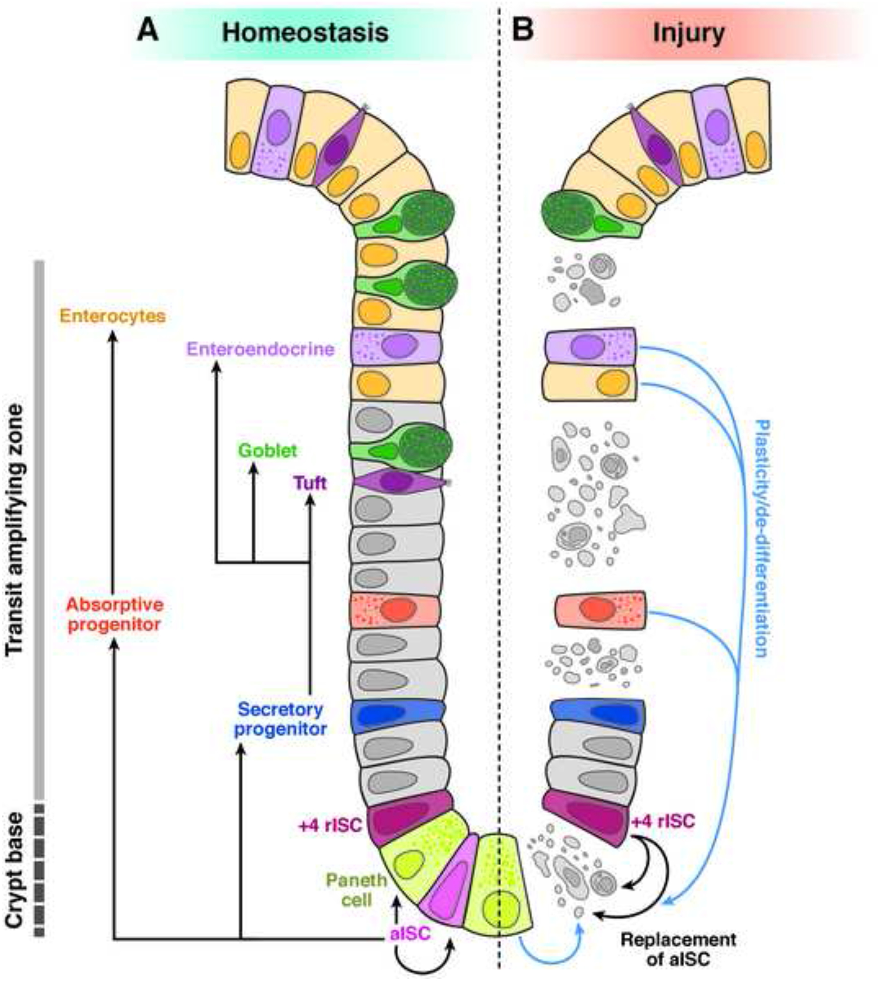
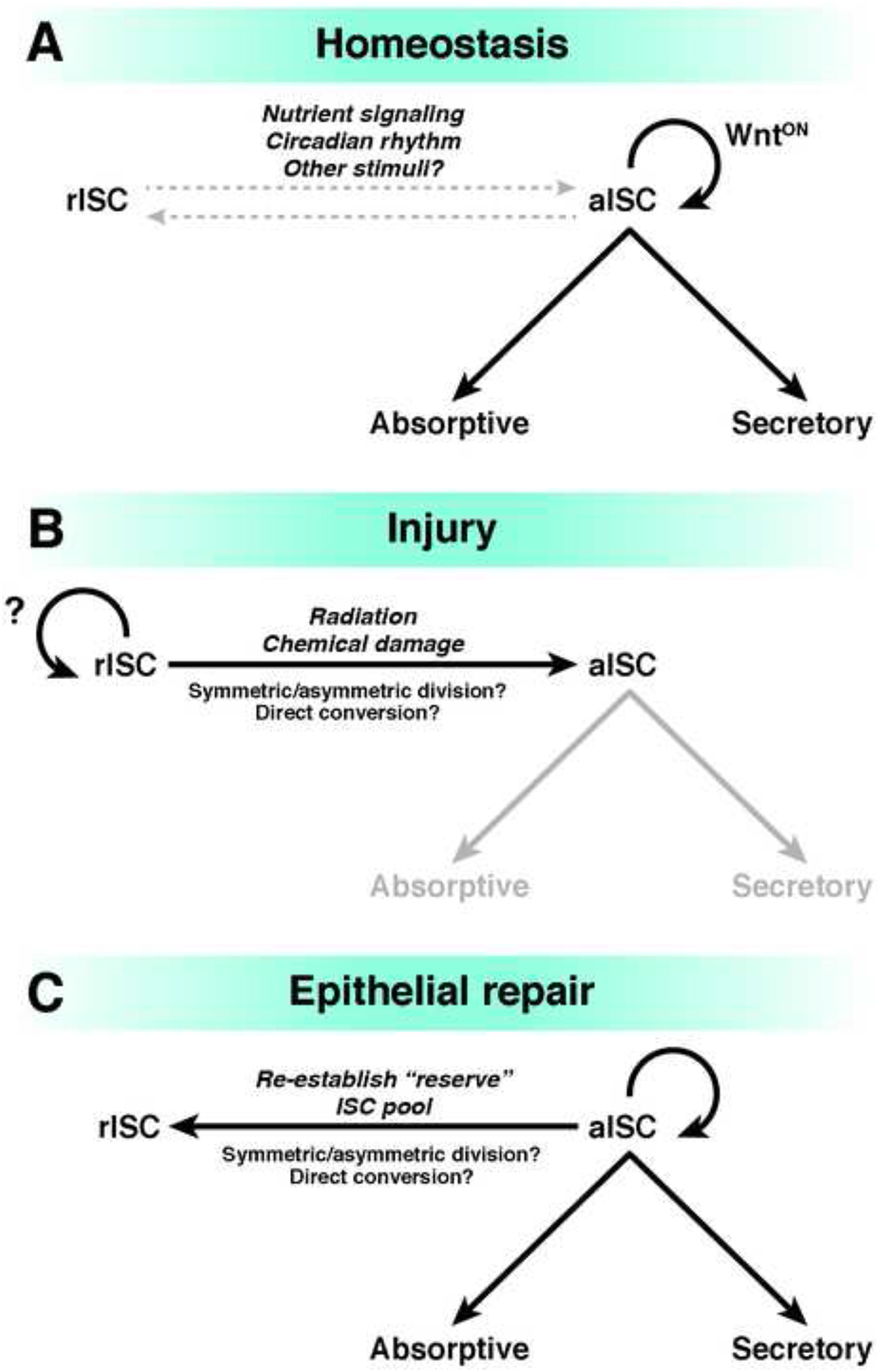
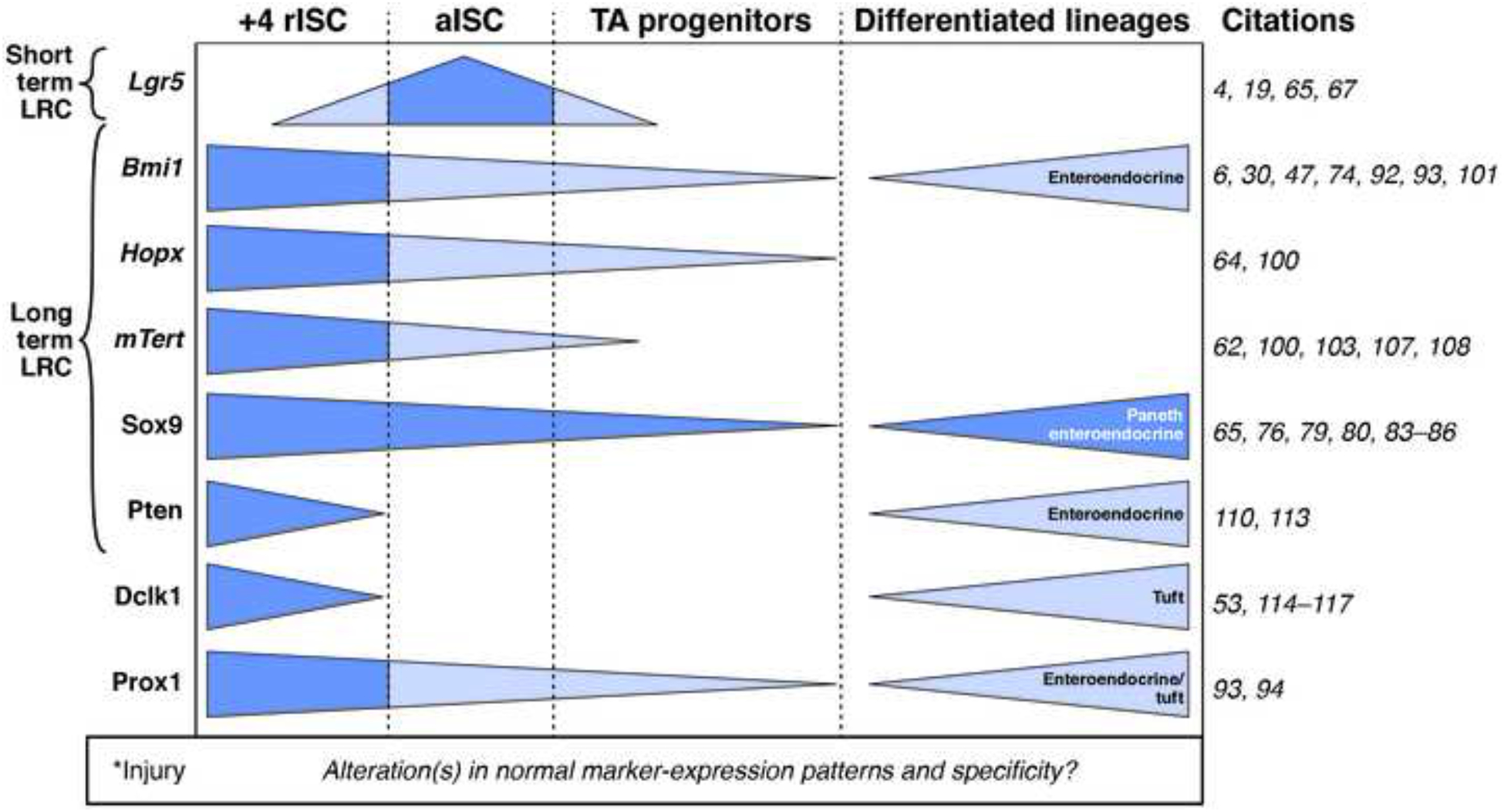
Renewal of the intestinal epithelium occurs approximately every week and requires a careful balance between cell proliferation and differentiation to maintain proper lineage ratios and support absorptive, secretory, and barrier functions. We review models used to study the mechanisms by which intestinal stem cells (ISCs) fuel the rapid turnover of the epithelium during homeostasis and might support epithelial regeneration after injury. In anatomically defined zones of the crypt stem cell niche, phenotypically distinct active and reserve ISC populations are believed to support homeostatic epithelial renewal and injury-induced regeneration, respectively. However, other cell types previously thought to be committed to differentiated states might also have ISC activity and participate in regeneration. Efforts are underway to reconcile the proposed relatively strict hierarchical relationships between reserve and active ISC pools and their differentiated progeny; findings from models provide evidence for phenotypic plasticity that is common among many if not all crypt-resident intestinal epithelial cells. We discuss the challenges to consensus on ISC nomenclature, technical considerations, and limitations inherent to methodologies used to define reserve ISCs, and the need for standardized metrics to quantify and compare the relative contributions of different epithelial cell types to homeostatic turnover and post-injury regeneration. Increasing our understanding of the high-resolution genetic and epigenetic mechanisms that regulate reserve ISC function and cell plasticity will help refine these models and could affect approaches to promote tissue regeneration after intestinal injury.
Keywords: Intestine; Regeneration; Stem Cells.
Copyright © 2018 AGA Institute. Published by Elsevier Inc. All rights reserved.
Conflict of interest statement
The authors declare no conflicts of interest
Figures
References
-
- Cells IP, Cheng H. Origin, Differentiation and Renewal of the Four Main Epithelial Cell Types in the Mouse Small Intestine . Dev. Dynamics 1974; 141:537–561. - PubMed
-
- Bjerknes M, Cheng H. The stem-cell zone of the small intestinal epithelium. I. Evidence from Paneth cells in the adult mouse. Am J Anat 1981;160:51–63. - PubMed
-
- Barker N, Es van JH, Kuipers J, et al. Identification of stem cells in small intestine and colon by marker gene Lgr5. Nature 2007;449:1003–1007. - PubMed
-
- Sato T, Vries RG, Snippert HJ, et al. Single Lgr5 stem cells build crypt–villus structures in vitro without a mesenchymal niche. Nature 2009;459:262–265. - PubMed
-
- Sato T, Clevers H. Growing Self-Organizing Mini-Guts from a Single Intestinal Stem Cell: Mechanism and Applications. Science (80−) 2013;340:1190–1194. - PubMed
Publication types
MeSH terms
Substances
Grants and funding
LinkOut - more resources
Full Text Sources
Other Literature Sources
Medical
Research Materials
