. 2018 Aug 17;37(1):196.
doi: 10.1186/s13046-018-0839-7.

IL-33 facilitates proliferation of colorectal cancer dependent on COX2/PGE2

Affiliations

IL-33 facilitates proliferation of colorectal cancer dependent on COX2/PGE2

Yongkui Li et al. J Exp Clin Cancer Res. .

Erratum in

Abstract

Background: Interleukin-33 (IL-33) participates in various types of diseases including cancers. Previous studies of this cytokine in cancers mainly focused on its regulation on immune responses by which IL-33 modulated cancer progression. The IL-33 triggered signals in cancer cells remain unclear.

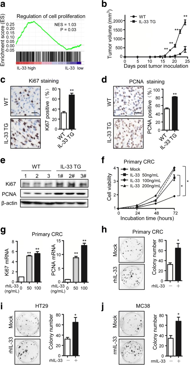
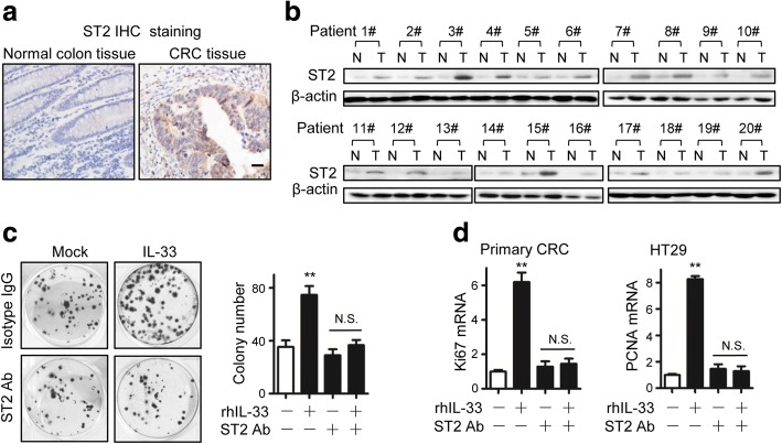
Methods: We analyzed IL-33 gene expression in human colorectal cancer (CRC) tissues and carried out gene enrichment analysis with TCGA Data Portal. We studied CRC proliferation in vivo by inoculating MC38 tumors in IL-33 transgenic mice. We investigated the cell proliferation in vitro with primary CRC cells isolated from fresh human CRC tissues, human CRC cell line HT-29 and mouse CRC cell line MC38. To evaluate the proliferation modulating effects of recombinant IL-33 incubation and other administrated factors, we measured tumor growth, colony formation, cell viability, and the expression of Ki67 and proliferating cell nuclear antigen (PCNA). We used several inhibitors, prostaglandin E2 (PGE2) neutralizing antibody, ST2 blocking antibody and specific shRNA expressing plasmid to study the pathway mediating IL-33-induced CRC proliferation. The IL-33 receptor ST2 in human CRC tissues was detected by immunohistochemistry staining and western blotting. The ST2-positive or negative subsets of primary CRC cells were acquired by flow cytometry sorting.

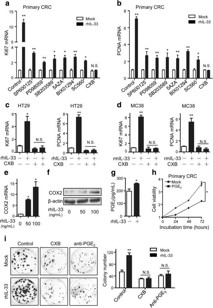
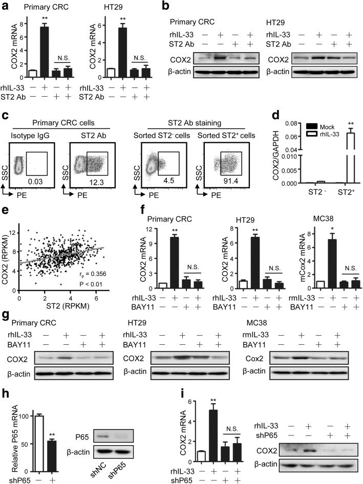
Results: We found that IL-33 expression was correlated with the gene signature of cell proliferation in 394 human CRC samples. The MC38 tumors grew more rapidly and the tumor Ki67 and PCNA were expressed at higher levels in IL-33 transgenic mice than in wild-type mice. IL-33 promoted cell growth, colony formation and expression of Ki67 and PCNA in primary CRC cells as well as CRC cell lines. IL-33 activated cycloxygenase-2 (COX2) expression and increased PGE2 production, whereas the COX2 selective inhibitor and PGE2 neutralizing antibody abolished the proliferation promoting effect of IL-33. ST2 blockade, ST2-negative sorting, NF-κB specific inhibitor and NF-κB specific shRNA (shP65) abrogated the COX2 induction caused by IL-33.

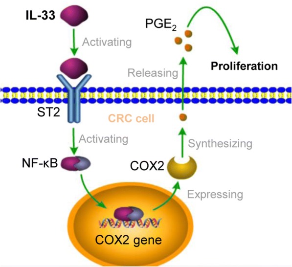
Conclusion: IL-33 facilitates proliferation of colorectal cancer dependent on COX2/PGE2. IL-33 functions via its receptor ST2 and upregulates COX2 expression through NF-κB signaling. Understanding the IL-33 signal transduction in CRC cells provides potential therapeutic targets for clinical treatment.

Keywords: COX2; Colorectal cancer; IL-33; PGE2; Proliferation.


Conflict of interest statement

Ethics approval and consent to participate

The experiments with animals and human samples in this study were conducted according to the principles of the Declaration of Helsinki and approved by the Institutional Review Boards of Tongji Medical College at Huazhong University of Science and Technology (Wuhan, Hubei, China).

Consent for publication

All patients signed informed consent for the publication of the clinical images and their clinical details.

Competing interests

The authors declare that they have no competing interests.

Publisher’s Note

Springer Nature remains neutral with regard to jurisdictional claims in published maps and institutional affiliations.

Figures

Fig. 1
Fig. 1
IL-33 promotes CRC proliferation both in vivo and in vitro. a Correlation between IL-33 transcripts and the genes involved in the regulation of cell proliferation in CRC. Gene set enrichment analysis was performed using CRC TCGA database. NES = 1.03, P = 0.03. b Growth curves of MC38 tumors inoculated in IL-33 transgenic mice (IL-33 TG) or wild-type mice (WT). n = 7. c, d Immunohistochemical staining of Ki67 (c) and PCNA (d) in the MC38 tumors recovered from wild-type and IL-33 transgenic mice at Day 22 post inoculation. The representative images and the statistical proportions of positive cells are shown. Scale bar, 50 μm. n = 7. Data expressed as mean ± SEM. **, P < 0.01. e Western blot of Ki67 and PCNA in the MC38 tumors recovered from wild-type and IL-33 transgenic mice. n = 3. f Cell viabilities of human primary CRC cells incubated with 0, 50, 100 or 200 ng/mL of rhIL-33 in medium at 24th, 48th and 72nd h. Six parallel wells were set for each treatment. The experiment was performed three times. Data expressed as mean ± SEM. * P < 0.05. g Ki67 and PCNA mRNA levels in primary CRC cells incubated with rhIL-33 (0, 50 or 100 ng/mL) for 24 h. Each experiment was performed three times. Three parallel wells were set for each treatment. Data expressed as mean ± SEM. ** P < 0.01. h, i, j The flat colony formation with 500 primary CRC cells (h) and 500 HT29 cells (i) incubated with rhIL-33 (100 ng/mL) and the flat colony formation with 500 MC38 cells (j) incubated with rmIL-33 (100 ng/mL). The number of colony was counted at Day 10. Each experiment was performed three times. Three parallel wells were set for each treatment. The representative images of colonies and the statistical data are shown. Data expressed as mean ± SEM. * P < 0.05
Fig. 2
Fig. 2
COX2/PGE2 mediates the proliferation promoting function of IL-33. a, b The relative mRNA levels of Ki67 (a) and PCNA (b) in primary CRC cells responding to rhIL-33 (100 ng/mL) incubation and/ or indicated inhibitors (SB203580, 10 μg/mL; PD98059, 20 μg/mL; SP600125, 10 μg/mL; BIX01294, 2 μM; 5Aza, 10 μM; SC560, 0.1 μg/mL; celecoxib, 20 μg/mL) for 24 h. c The relative mRNA levels of Ki67 and PCNA in HT-29 cells incubated with rhIL-33 (100 ng/mL) or/ and celecoxib (CXB) (20 μg/mL) in medium for 24 h. d The relative mRNA levels of Ki67 and PCNA in MC38 cells incubated with rmIL-33 (100 ng/mL) or/ and celecoxib (CXB) (20 μg/mL) in medium for 24 h. e, f The mRNA (e) and protein (f) expression of COX2 in primary CRC cells incubated with 0, 50 or 100 ng/mL of rhIL-33 in medium for 24 h. g PGE2 concentrations in the supernatants of primary CRC cells incubated with rhIL-33-contained RPMI medium or blank RPMI medium for 48 h. h Cell viabilities of primary CRC cells incubated with or without PGE2 (50 ng/mL) in medium. i The flat colony formation of primary CRC cells incubated for 15 days in medium containing different factors as indicated (IL-33, 100 ng/mL; celecoxib, 20 μg/mL; anti-PGE2, 2 μg/mL). The representative images of colonies and the statistical data are shown. Three parallel wells were set for each treatment. Each experiment was performed three times. Data expressed as mean ± SEM. * P < 0.05. ** P < 0.01
Fig. 3
Fig. 3
IL-33 facilitated CRC proliferation by signaling its receptor ST2. a Immunohistochemical staining of ST2 in the CRC tissues and the adjacent normal colon tissues (20 pairs). The representative images are shown. Scale bar, 20 μm. b ST2 expression levels in the paired CRC tissues (T) and the adjacent normal colon tissues (N) analyzed by Western blotting. c The flat colony formation of primary CRC cells incubated for 15 days in RPMI medium or RPMI medium containing rhIL-33 (100 ng/mL) or/ and ST2 antibody (2 μg/mL). Three parallel wells were set for each treatment. Each experiment was performed three times. The representative images of colonies and the statistical data are shown. Data expressed as mean ± SEM. ** P < 0.01. d Ki67 and PCNA expression in primary CRC cells responding to the incubation with rhIL-33 (100 ng/mL) or/ and ST2 antibody (2 μg/mL) for 24 h. Three parallel wells were set for each treatment. Each experiment was performed three times. Data expressed as mean ± SEM. ** P < 0.01
Fig. 4
Fig. 4
IL-33/ST2 upregulates COX2 expression through NF-κB signaling. a,b The COX2 mRNA (a) and protein (b) expression in primary CRC cells or HT29 cells responding to the incubation with rhIL33 (100 ng/mL) or/ and ST2 antibody (2 μg/mL) for 24 h. Each experiment was performed three times. Data expressed as mean ± SEM. ** P < 0.01. c ST2 expression distribution in primary CRC cells, sorted ST2-negative and sorted ST2-positive primary CRC cells. The proportion of ST2 positive subset is shown. d Relative COX2 mRNA levels in ST2-negative or ST2-positive primary CRC cells responding to IL-33 (100 ng/mL) incubation for 24 h in 24-well plates (1 × 105 cells per well). Three parallel wells were set for each treatment. Data expressed as mean ± SEM. ** P < 0.01. e The correlation between COX2 and ST2 transcripts in 394 CRC samples recorded in TCGA database. These two sets of data both have a normal distribution. Pearson r = 0.356, P < 0.01. f COX2 mRNA levels in primary CRC cells, HT29 cells and MC38 responding to the incubation with IL-33 (100 ng/mL) or/ and BAY11–7082 (10 μM). Three parallel wells were set for each treatment. Each experiment was performed three times. Data expressed as mean ± SEM. * P < 0.05. ** P < 0.01. g COX2 protein levels in primary CRC cells, HT29 cells and MC38 responding to the incubation with IL-33 (100 ng/mL) or/ and BAY11–7082 (10 μM). Each experiment was performed three times. h The knocking-down efficiency of NF-κB P65 in HT29 cells. The P65 mRNA (left panel) and protein (right panel) were both detected. Data expressed as mean ± SEM. ** P < 0.01. i COX2 mRNA (left panel) and protein (right panel) levels responding to IL-33 incubation (100 ng/mL) for 24 h in HT29 cells transfected with short hairpin RNA expressing plasmid against NF-κB P65 (shP65) or nonsense RNA expressing plasmid (shNC). Each experiment was performed three times. Data expressed as mean ± SEM. ** P < 0.01
Fig. 5
Fig. 5
The pathway through which IL-33 stimulates the proliferation signaling of CRC cells

Similar articles

Cited by

References

    1. Liew FY, Pitman NI, McInnes IB. Disease-associated functions of IL-33: the new kid in the IL-1 family. Nat Rev Immunol. 2010;10(2):103–110. doi: 10.1038/nri2692. - DOI - PubMed
    1. Schmitz J, Owyang A, Oldham E, Song Y, Murphy E, McClanahan TK, Zurawski G, Moshrefi M, Qin J, Li X, et al. IL-33, an interleukin-1-like cytokine that signals via the IL-1 receptor-related protein ST2 and induces T helper type 2-associated cytokines. Immunity. 2005;23(5):479–490. doi: 10.1016/j.immuni.2005.09.015. - DOI - PubMed
    1. Saigusa R, Asano Y, Taniguchi T, Hirabayashi M, Nakamura K, Miura S, Yamashita T, Takahashi T, Ichimura Y, Toyama T, et al. Fli1-haploinsufficient dermal fibroblasts promote skin-localized transdifferentiation of Th2-like regulatory T cells. Arthritis research & therapy. 2018;20(1):23. doi: 10.1186/s13075-018-1521-3. - DOI - PMC - PubMed
    1. Fang M, Li Y, Huang K, Qi S, Zhang J, Zgodzinski W, Majewski M, Wallner G, Gozdz S, Macek P, et al. IL33 promotes Colon Cancer cell Stemness via JNK activation and macrophage recruitment. Cancer Res. 2017;77(10):2735–2745. doi: 10.1158/0008-5472.CAN-16-1602. - DOI - PMC - PubMed
    1. Dinarello CA. An IL-1 family member requires caspase-1 processing and signals through the ST2 receptor. Immunity. 2005;23(5):461–462. doi: 10.1016/j.immuni.2005.10.004. - DOI - PubMed