. 2018 Sep 28;293(39):15304-15315.
doi: 10.1074/jbc.RA118.004444. Epub 2018 Aug 17.

Biochemical properties and in planta effects of NopM, a rhizobial E3 ubiquitin ligase

Affiliations

Biochemical properties and in planta effects of NopM, a rhizobial E3 ubiquitin ligase

Chang-Chao Xu et al. J Biol Chem. .

Abstract

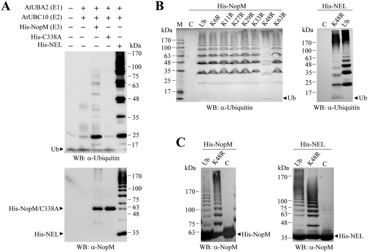
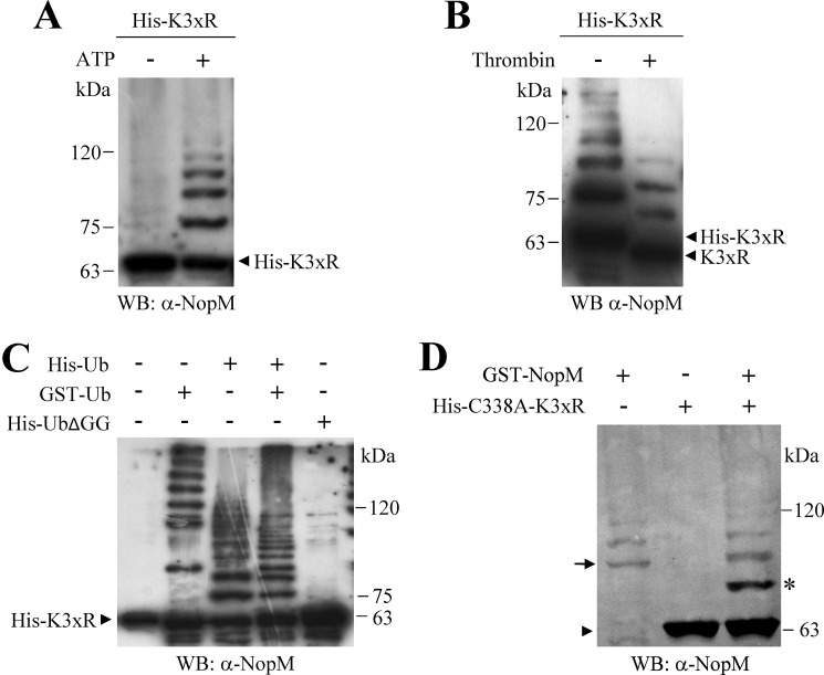
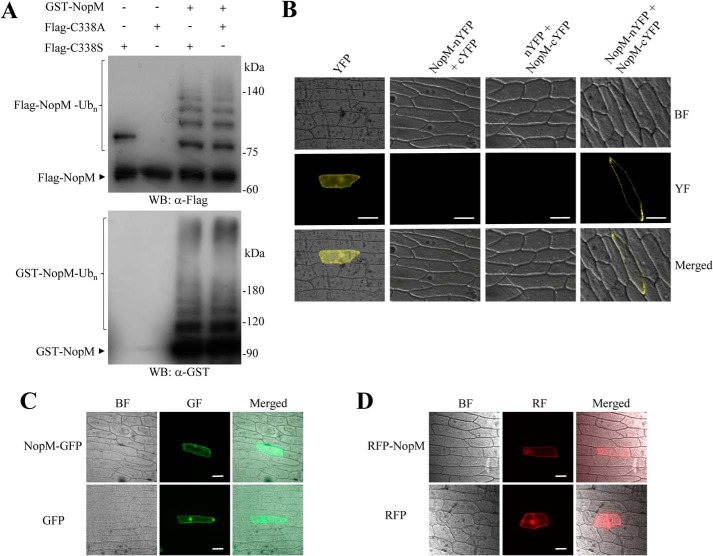
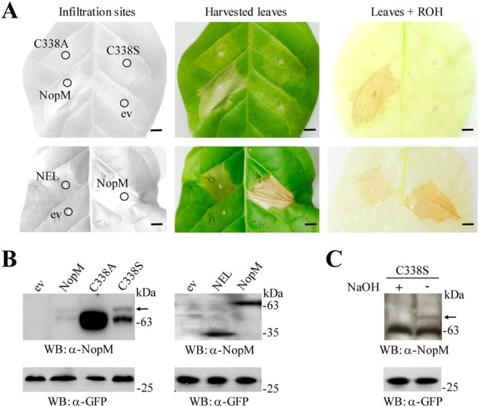
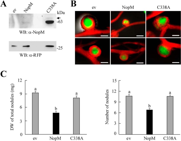
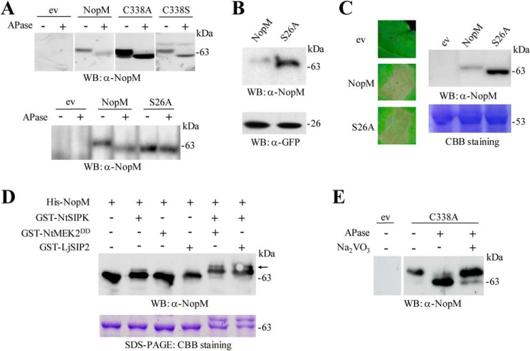
Nodulation outer protein M (NopM) is an IpaH family type three (T3) effector secreted by the nitrogen-fixing nodule bacterium Sinorhizobium sp. strain NGR234. Previous work indicated that NopM is an E3 ubiquitin ligase required for an optimal symbiosis between NGR234 and the host legume Lablab purpureus Here, we continued to analyze the function of NopM. Recombinant NopM was biochemically characterized using an in vitro ubiquitination system with Arabidopsis thaliana proteins. In this assay, NopM forms unanchored polyubiquitin chains and possesses auto-ubiquitination activity. In a NopM variant lacking any lysine residues, auto-ubiquitination was not completely abolished, indicating noncanonical auto-ubiquitination of the protein. In addition, we could show intermolecular ubiquitin transfer from NopM to C338A (enzymatically inactive NopM form) in vitro Bimolecular fluorescence complementation analysis provided clues about NopM-NopM interactions at plasma membranes in planta NopM, but not C338A, expressed in tobacco cells induced cell death, suggesting that E3 ubiquitin ligase activity of NopM induced effector-triggered immunity responses. Likewise, expression of NopM in Lotus japonicus caused reduced nodule formation, whereas expression of C338A showed no obvious effects on symbiosis. Further experiments indicated that serine residue 26 of NopM is phosphorylated in planta and that NopM can be phosphorylated in vitro by salicylic acid-induced protein kinase (NtSIPK), a mitogen-activated protein kinase (MAPK) of tobacco. Hence, NopM is a phosphorylated T3 effector that can interact with itself, with ubiquitin, and with MAPKs.

Keywords: E3 ubiquitin ligase; bacteria; effector protein; phosphorylation; symbiosis; type III secretion system (T3SS); ubiquitination.


Conflict of interest statement

The authors declare that they have no conflicts of interest with the contents of this article

Figures

Figure 1.
Figure 1.
Ubiquitination reactions catalyzed by NopM and variants. A, Western blot (WB) analysis of ubiquitination reactions with NopM and the variants C338A (no activity) and NEL (increased activity). Recombinant E1, E2, and Ub proteins of A. thaliana were used for the reactions. Western blottings were developed with anti-ubiquitin (top) or anti-NopM (bottom) antibodies. B and C, ubiquitination reactions catalyzed by NopM or NEL with ubiquitin or indicated ubiquitin variants with lysine to arginine substitutions. Control reactions without ubiquitin are also shown (lanes C). Western blot analysis was performed with anti-ubiquitin (B) or anti-NopM (C) antibodies. Lane M, molecular weight markers.
Figure 2.
Figure 2.
K3xR variant shows auto-ubiquitination activity. A–C, His-tagged K3xR protein (NopM with K502R, K531R, and K537R substitutions) was used for ubiquitination reactions (2 h), and auto-ubiquitination was detected with the anti-NopM antibody. Nonubiquitinated K3xR is marked by arrowheads. A, reactions (2 h) were performed with (+) or without (−) ATP. B, incubation of reaction products with and without thrombin (removal of the N-terminal His tag). C, reactions with GST-tagged ubiquitin, His-tagged ubiquitin, with a mixture (Ub mix; GST- and His-tagged ubiquitin, 1:1), or with UbΔGG (Ub without C-terminal di-glycine residues). D, analysis of intermolecular transfer of His-tagged ubiquitin from GST–NopM to His-tagged K3xR-C338A (NopM with C338A, K502R, K531R, and K537R substitutions). Nonubiquitinated His–K3xR–C338A is marked by an arrowhead, mono-ubiquitinated His–K3xR–C338A by an asterisk, and GST–NopM by an arrow. WB, Western blot.
Figure 3.
Figure 3.
Analysis of NopM–NopM interactions and subcellular localization of NopM fused to fluorescent proteins. A, analysis of intermolecular transfer of ubiquitin in vitro. The Flag-tagged NopM variants C338A (enzymatically inactive) and C338S (forming a mono-ubiquitinated conjugate) were ubiquitinated by GST–NopM. Western blot (WB) were probed with anti-Flag or anti-GST antibodies. B, BiFC analysis of NopM–NopM interactions in vivo. Onion cells expressing indicated protein combinations were microscopically analyzed for yellow fluorescence (YF) emission and under bright field (BF) illumination. Co-expression of NopM–nYFP with NopM–cYFP resulted in formation of a BiFC complex at plasma membranes. Bars, 100 μm. C and D, subcellular localization of full-length NopM fusion proteins. Fluorescent NopM proteins with C-terminal GFP tag (C) or with N-terminal RFP tag (D) were expressed in onion cells. GFP and RFP alone were expressed for comparison. Emission of green fluorescence (GF), red fluorescence (RF), and bright field conditions were used for microscopic examination. Bars, 100 μm.
Figure 4.
Figure 4.
Expression of NopM and variants in tobacco. A, photographs of tobacco leaves expressing NopM, NEL, C338A, and C338S, respectively. A. tumefaciens carrying the empty vector (ev) was used as a negative control. Photographs were taken 3 days after infiltration of the bacterial suspensions. Harvested leaves were decolorized by boiling in ethanol (ROH) to visualize necrotic tissue. Bars, 0.5 cm. B, Western blot (WB) analysis of expressed proteins. Crude proteins from transformed tobacco leaves were isolated 2 days after infiltration with agrobacteria. Western blots were performed with 5 μl of the prepared extract and anti-NopM or anti-GFP antibodies. The upper bands (above NopM and C338S; marked by an arrow) likely represent mono-ubiquitinated forms. C, treatment of extracted C338S with 100 mm NaOH (final concentration) resulted in disappearance of the upper band, suggesting ubiquitination on the serine residue by formation of an oxyester bond.
Figure 5.
Figure 5.
Expression of NopM and C338A in L. japonicus roots. Agrobacterium rhizogenes-mediated transformation was used to obtain transgenic roots expressing NopM or the enzymatically inactive variant C338A. Plants transformed with the empty vector (ev) containing an RFP expression cassette were used as a negative control. A, proteins from transgenic roots were isolated 30 days after transformation, and 5 μl of extracts were used for Western blot (WB) analysis with anti-NopM and anti-RFP antibodies. B, examples of pink nodules formed on the transformed L. japonicus roots 25 days after inoculation with M. loti MAFF303099 (GFP). Transgenic roots show RFP expression (red fluorescence). Green fluorescence indicates the presence of bacteria within nodules. Bars, 500 μm. C, quantification of nodule biomass (dry weight (DW)) and number of formed nodules per plant. Data indicate mean ± S.E. (n = 10). NopM-expressing roots formed significantly fewer nodules (different letters indicate differences; Kruskal-Wallis test, p < 0.05).
Figure 6.
Figure 6.
Phosphorylation of NopM in planta and in vitro. A, analysis of NopM and indicated variants isolated from tobacco leaves transformed with A. tumefaciens. Agrobacteria carrying the empty vector (ev) were used as negative control. Soluble proteins were extracted from the transformed leaf tissue, and aliquots were treated with APase. Samples were then subjected to Western blot (WB) analysis with anti-NopM antibodies. B, Western blot analysis of NopM and the S26A variant expressed in tobacco leaves. For comparison, co-expressed GFP was also analyzed in the obtained protein extracts. C, expression of the S26A variant in tobacco induces cell death. NopM expression and empty vector controls were used for comparison. The pictures were taken 3 days after infiltration with agrobacteria. A corresponding Western blot confirmed expression of the proteins. Coomassie Brilliant Blue (CBB) staining of ribulose-bisphosphate carboxylase/oxygenase large subunit in a parallel gel indicated the use of equal amounts of proteins. D, in vitro phosphorylation of NopM by NtSIPK. Indicated proteins with GST or His tags were expressed in E. coli and purified by affinity chromatography. The MAPK kinases NtMEK2DD and LjSIP2 were used for activation of NtSIPK. Proteins were incubated in phosphorylation buffer containing ATP for 30 min at 30 °C. The samples were analyzed by Western blotting using an anti-NopM antibody. NopM phosphorylated by activated NtSIPK migrated more slowly on the gel (upper bands marked by an arrow). A parallel protein gel was stained with Coomassie Brilliant Blue. E, analysis of the C338A variant expressed in L. japonicus roots. Soluble proteins were extracted 28 days after transformation, and aliquots were treated with APase and 10 mm Na3VO4. Samples were then subjected to Western blot analysis with anti-NopM antibodies.

Similar articles

Cited by

References

    1. Galán J. E., Lara-Tejero M., Marlovits T. C., and Wagner S. (2014) Bacterial type III secretion systems: specialized nanomachines for protein delivery into target cells. Annu. Rev. Microbiol. 68, 415–438 10.1146/annurev-micro-092412-155725 - DOI - PMC - PubMed
    1. Marín M., Uversky V. N., and Ott T. (2013) Intrinsic disorder in pathogen effectors: protein flexibility as an evolutionary hallmark in a molecular arms race. Plant Cell 25, 3153–3157 10.1105/tpc.113.116319 - DOI - PMC - PubMed
    1. Büttner D. (2016) Behind the lines–actions of bacterial type III effector proteins in plant cells. FEMS Microbiol. Rev. 40, 894–937 10.1093/femsre/fuw026 - DOI - PMC - PubMed
    1. Cui H., Tsuda K., and Parker J. E. (2015) Effector-triggered immunity: from pathogen perception to robust defense. Annu. Rev. Plant Biol. 66, 487–511 10.1146/annurev-arplant-050213-040012 - DOI - PubMed
    1. Staehelin C., and Krishnan H. B. (2015) Nodulation outer proteins: double-edged swords of symbiotic rhizobia. Biochem. J. 470, 263–274 10.1042/BJ20150518 - DOI - PubMed

Publication types

MeSH terms

Substances

LinkOut - more resources