

Magnetic resonance imaging in Alzheimer's disease and mild cognitive impairment
- PMID: 30120563
- PMCID: PMC6517561
- DOI: 10.1007/s00415-018-9016-3
Magnetic resonance imaging in Alzheimer's disease and mild cognitive impairment
Abstract
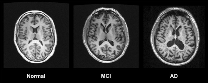
Research utilizing magnetic resonance imaging (MRI) has been crucial to the understanding of the neuropathological mechanisms behind and clinical identification of Alzheimer's disease (AD) and mild cognitive impairment (MCI). MRI modalities show patterns of brain damage that discriminate AD from other brain illnesses and brain abnormalities that are associated with risk of conversion to AD from MCI and other behavioural outcomes. This review discusses the application of various MRI techniques to and their clinical usefulness in AD and MCI. MRI modalities covered include structural MRI, diffusion tensor imaging (DTI), arterial spin labelling (ASL), magnetic resonance spectroscopy (MRS), and functional MRI (fMRI). There is much evidence supporting the validity of MRI as a biomarker for these disorders; however, only traditional structural imaging is currently recommended for routine use in clinical settings. Future research is needed to warrant the inclusion for more advanced MRI methodology in forthcoming revisions to diagnostic criteria for AD and MCI.
Keywords: Alzheimer’s disease; Magnetic resonance imaging; Mild cognitive impairment; Neuropathology.
Conflict of interest statement
The authors of this manuscript have no conflicts of interests to disclose relevant to the current review.
Figures

References
-
- Harrington CR. The molecular pathology of Alzheimer’s disease. Neuroimaging Clin N Am. 2012;22:11–22. - PubMed
-
- McKhann GM, Knopman DS, Chertkow H, Hyman BT, Jack CR, Kawas CH, Klunk WE, Koroshetz WJ, Manly JJ, Mayeux R. The diagnosis of dementia due to Alzheimer’s disease: recommendations from the National Institute on Aging-Alzheimer’s Association workgroups on diagnostic guidelines for Alzheimer’s disease. Alzheimer’s Dement. 2011;7:263–269. - PMC - PubMed
-
- Albert MS, DeKosky ST, Dickson D, Dubois B, Feldman HH, Fox NC, Gamst A, Holtzman DM, Jagust WJ, Petersen RC. The diagnosis of mild cognitive impairment due to Alzheimer’s disease: recommendations from the National Institute on Aging-Alzheimer’s Association workgroups on diagnostic guidelines for Alzheimer’s disease. Alzheimer’s Dement. 2011;7:270–279. - PMC - PubMed
-
- Politis M, Piccini P. Positron emission tomography imaging in neurological disorders. J Neurol. 2012;259:1769–1780. - PubMed
-
- Braak H, Braak E. Neuropathological stageing of Alzheimer-related changes. Acta Neuropathol. 1991;82:239–259. - PubMed
Publication types
MeSH terms
Grants and funding
LinkOut - more resources
Full Text Sources
Other Literature Sources
Medical
